Introduction To Clinical Context And Proposed Utility Of Modality
MRI became the method of choice for detection and staging of prostate cancer . Adapted from breast imaging a Prostate Imaging Reporting and Data System was published by the European Society of Urogenital Radiology : PI-RADS version 1 . This first guideline paper was based on a summary score for each lesion assessed in different sequences of mpMRI, consisting of T2w, DWI and DCE-MRI and spectroscopy facultatively. These guidelines have been updated recently by a steering committee including the American College of Radiology , ESUR and the AdMeTech Foundation to the PI-RADS v2 . In this version spectroscopy was omitted and DCE-MRI was attributed a minor role. In contrast to version 1 each lesion is attributed a single score based on findings of mpMRI. The objectives of these guidelines were to promote global standardisation of prostate imaging, to improve detection, localisation, characterisation, risk stratification of prostate cancer in treatment naïve prostate as well as to improve communication with referring urologists. The latest PI-RADS version assesses the likelihood of clinically significant prostate cancer on a 5-point scale for each lesion as follows:
-
PI-RADS 1 Very low
-
PI-RADS 2 Low
-
PI-RADS 3 Intermediate
-
PI-RADS 4 High
-
PI-RADS 5 Very high
For corresponding examples of findings see Fig. .
Fig. 1
Staging Of T3a Disease
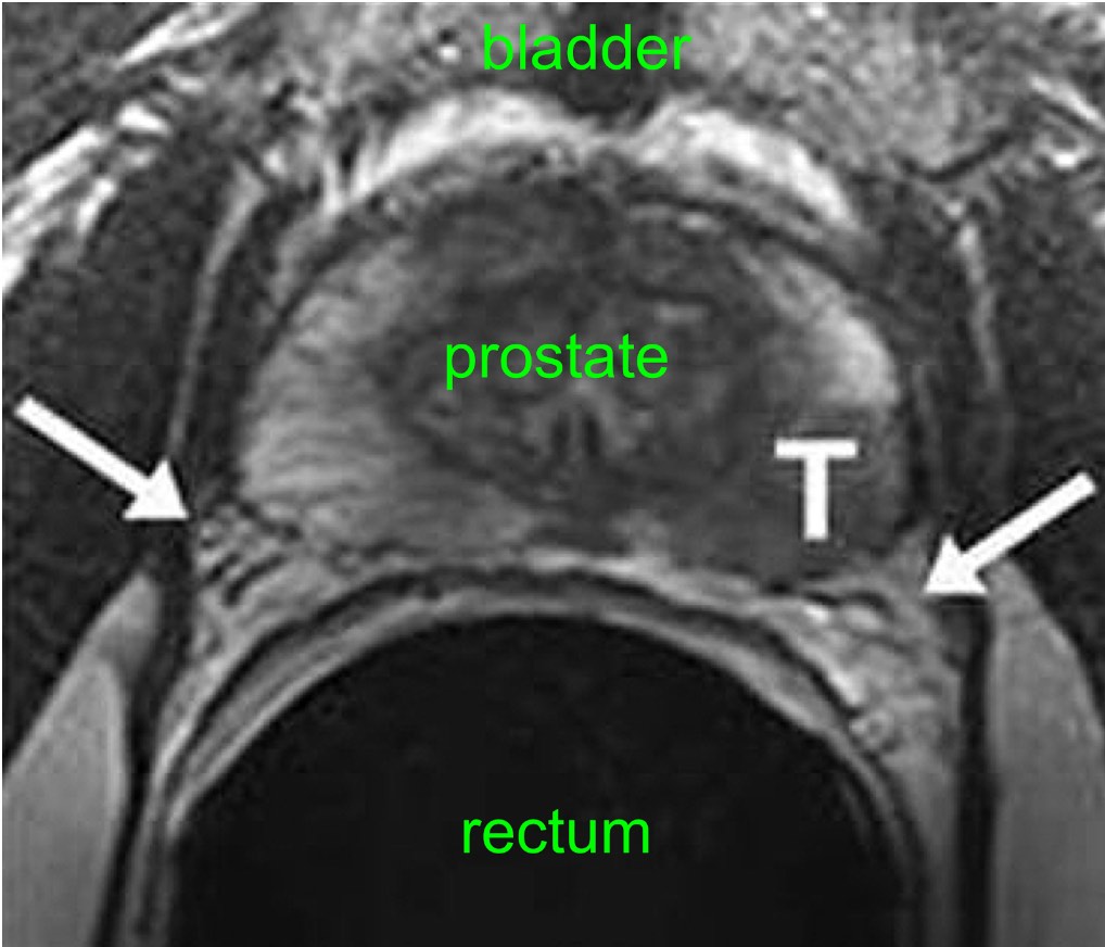
A 2016 meta-analysis of 75 total studies found that pooled data for EPE showed sensitivity and specificity of 57% and 91% respectively, concluding that MRI has high specificity but poor sensitivity for local prostate cancer staging .40 EPE sensitivity was not improved by endorectal coil use, but a sub-analysis of higher field strengths , showed improved sensitivity of 68%,40 demonstrating a potential area for improvement.
|
Table 2 Summary of MRI Performance Characteristics for Detection of T3a, T3b and T4 Disease |
Length of tumor contact at capsule can be a particularly helpful measurement when determining risk of EPE, which can be seen by its inclusion in both aforementioned systems, however there is still lack of data about its predictive value. Previous studies indicate length of contact has good accuracy and inter-reader agreement. Rosenkrantz et al reported that inter-reader agreement was 0.7 for assessments based on length of contact and 0.490.59 for subjective assessments.46 There remains disagreement on the exact threshold to use for this criterion. The PIRADS v2 guidelines recommend using > 10mm, while other sources vary from 620mm.47 Factors such as technical method of measurement and pretest probability can, in part, explain this variation. One study found that for high-grade tumors , positive predictive value of 5 mm of contact was 90.9% versus positive predictive value of 90.4% for 12.5 mm for lower grade tumors.45
Use In Men Already Diagnosed With Prostate Cancer
The PSA test can also be useful if you have already been diagnosed with prostate cancer.
- In men just diagnosed with prostate cancer, the PSA level can be used together with physical exam results and tumor grade to help decide if other tests are needed.
- The PSA level is used to help determine the stage of your cancer. This can affect your treatment options, since some treatments are not likely to be helpful if the cancer has spread to other parts of the body.
- PSA tests are often an important part of determining how well treatment is working, as well as in watching for a possible recurrence of the cancer after treatment .
Read Also: What Does Prostate Inflammation Feel Like
Comparison Of Clinical And Mri Parameters Among Biopsy Isup Grade Groups
In 187 of all 200 patients, a PCA suspicious IL was found on mpMRI. In 13 patients, PCA was indistinct or masked. In ISUP 1, only 40/50 of the PCA were visible in mpMRI, whereas all PCA were visible in ISUP 4 and 5. 39/50 of the IL in ISUP 1 were localized in the peripheral zone, 35/50 in ISUP 2, 39/50 in ISUP 3, and 39/50 in ISUP 4 and 5. 7/50 of the IL in ISUP 1 were localized in the transition zone , and 4/50 of the IL in ISUP 1 were localized in the anterior stroma . Except for PCA diameter , ANOVA analysis showed significant differences for the means of all examined clinical and MRI-based parameters among the different ISUP grade groups with p=0.004 for signal increase on high b-value images, p=0.044 for ADC values using ss-EPI , and p< 0.001 for all others, respectively .
Will The Mri Be Done With An Endorectal Coil Or An External Pelvic Coil

Some radiology practices use an endorectal coil a probe-like device covered with latex which is inserted into the rectum and helps provide high-quality images of the prostate. With a newer, high-quality MRI system, endorectal coils are not necessary and an external pelvic coil can be used instead, eliminating patient discomfort while maintaining high quality images.
Read Also: Types Of Pet Scans For Prostate Cancer
Mri For Prostate Cancer Grading And Staging
Doctors can use prostate MRI tests to help stage prostate cancer. Urologists may refer men to radiology for a prostate MRI after a positive biopsy. The MRI test provides information that a biopsy might not, such as determining whether or not the cancer had spread beyond the prostate glandA gland is an organ that produces and secretes a chemical su… Full Definition.
This information, along with biopsy findings, PSA test results, and digital rectal exam results, can help the care team identify the best treatment options. For example:
- MRI images can determine with very good confidence whether or not the cancer has spread beyond the prostate gland. If it has, surgery may not be a good option, except in certain situations. BrachytherapyA form of radiotherapy where small radioactive implants are … Full Definition may not be a good option either, unless combined with other types of radiationHigh-energy particles that cause ionization and tissue damag… Full Definition therapy.
- MRI images can show the location of a tumorA lump or swelling caused by abnormal growth of cells. Not a… Full Definition. A mans tumor may be small but if it is close to the edge of the prostate, perhaps near the nerves important in erectile function or near the urethraThe duct urine passes through on the way… Full Definition or rectal walls, doctors might encourage the man to pursue treatment rather than active surveillance.
Continued Below
Current Lymph Node Evaluation
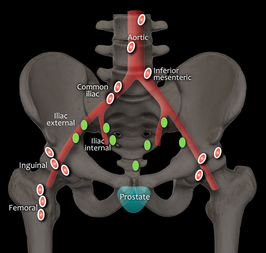
A designation of N1 disease comprises a positive lymph node in the obturator, external and internal, or pre-sacral iliac node bundles, which are considered the regional nodes. Presence of disease in lymph nodes outside of these four regions upstages the diagnosis to M1. In patients undergoing pre-biopsy MRI, PI-RADS v2 recommends a dedicated sequence for evaluation of lymph nodes that includes extension of study area to level of aortic bifurcation to fully incorporate evaluation of these regional nodal sites.
The detection of abnormal lymph nodes on MRI is currently limited to size, morphology and shape, and enhancement pattern.30 Other suggested evaluation criteria include size > 8mm , loss of fatty hilum, rounded shape, low T2W signal relative to primary tumor, and irregular border.61 Size alone is an unhelpful criterion for assessment of positive lymph node disease as there is significant overlap between positive and negative nodes, as well as reactive inflammatory changes from unrelated conditions which can produce false positive results. Studies have shown that the majority of positive lymph nodes dissected in extended-PLND following MRI have a short axis diameter of less than 5mm, below the aforementioned cutoff of 8mm.63 Further, smaller lymph nodes can harbor micro-metastasis that do not affect node size, leading to incorrect staging.64
Read Also: What Happens When You Remove Your Prostate
Understanding Your Mri Results
A doctor who specialises in imaging scans gives the MRI scan a score based on the results. This score helps your doctor decide on the next step and whether you need to have a biopsy. This is where your doctor takes a sample of your prostate gland.
This score is called the Likert or PI-RAD system. It estimates the risk that an area seen on the MRI scan may be a cancer or not. The score is from 1 to 5.
| Likert score | |
|---|---|
| Its highly unlikely that you have prostate cancer | |
| 2 | Its unlikely that you have prostate cancer |
| 3 | Its difficult to tell from the scan if a prostate cancer is present or not |
| 4 | Its likely that you have prostate cancer |
| 5 | Its very likely that you have prostate cancer |
What Does An Mri Scan Involve
Before the scan the doctor or nurse will ask questions about your health. As the scan uses magnets, they will ask whether you have any implants that could be attracted to the magnet. For example, if you have a pacemaker for your heart you may not be able to have an MRI scan. Youll also need to take off any jewellery or metal items.
You will lie very still on a table, which will move slowly into the scanner. MRI scanners are shaped like a doughnut or a long tunnel. If you dont like closed or small spaces , tell your radiographer .
The radiographer might give you an injection of a dye during the scan. The dye helps them see the prostate and other organs more clearly on the scan. It is usually safe, but can sometimes cause problems if you have kidney problems or asthma. So let the radiographer know if you have either of these, or if you know youre allergic to the dye or have any other allergies.
The scan takes 30 to 40 minutes. The machine wont touch you but it is very noisy and you might feel warm. The radiographer will leave the room but youll be able to speak to them through an intercom, and you might be able to listen to music through headphones.
Recommended Reading: When Should You Test For Prostate Cancer
Lymph Node Biopsy As A Separate Procedure
A lymph node biopsy is rarely done as a separate procedure. Its sometimes used when a radical prostatectomy isnt planned , but when its still important to know if the lymph nodes contain cancer.
Most often, this is done as a needle biopsy. To do this, the doctor uses an image to guide a long, hollow needle through the skin in the lower abdomen and into an enlarged node. The skin is numbed with local anesthesia before the needle is inserted to take a small tissue sample. The sample is then sent to the lab and looked at for cancer cells.
Correlation And Predictive Power Of The Isup Grade Group Among Clinical And Mri Parameters
All clinical parameters and almost all mpMRI parameters showed a significant correlation with the PCA ISUP grade group with the strongest association for the parameters MRI grading group and ADC values . No significant correlation was found for PCA diameter . The PI-RADS scoring system also correlated significantly with the ISUP grade groups .
Read Also: Can You Survive Aggressive Prostate Cancer
Tests To Diagnose And Stage Prostate Cancer
Most prostate cancers are first found as a result of screening. Early prostate cancers usually dont cause symptoms, but more advanced cancers are sometimes first found because of symptoms they cause.
If prostate cancer is suspected based on results of screening tests or symptoms, tests will be needed to be sure. If youre seeing your primary care doctor, you might be referred to a urologist, a doctor who treats cancers of the genital and urinary tract, including the prostate.
The actual diagnosis of prostate cancer can only be made with a prostate biopsy .
On this page
What Gets Stored In A Cookie

This site stores nothing other than an automatically generated session ID in the cookie no other information is captured.
In general, only the information that you provide, or the choices you make while visiting a web site, can be stored in a cookie. For example, the site cannot determine your email name unless you choose to type it. Allowing a website to create a cookie does not give that or any other site access to the rest of your computer, and only the site that created the cookie can read it.
Don’t Miss: Does Flomax Reduce Prostate Size
Genetic Testing For Some Men With Prostate Cancer
Some doctors now recommend that some men with prostate cancer be tested to look for certain inherited gene changes. This includes men in whom a family cancer syndrome is suspected, as well as men with prostate cancer that has certain high-risk features or that has spread to other parts of the body. Talk to your doctor about the possible pros, cons, and limitations of such testing.
Understanding Your Biopsy Results
A specialist doctor called pathologist looks at the prostate samples under a microspore. The results usually take about 2 to 4 weeks.
You might have a negative biopsy. This means that no cancer cells were found. Your doctor might recommend another biopsy even if the first was negative. They’ll discuss this with you. This is because in some cases biopsies can miss cancer.
A positive biopsy means that they have found cancer cells. A pathologist then grades each sample of prostate cancer cells based on how quickly they are likely to grow or how aggressive the cells look. You may hear this being called the Gleason score or Grade Group.
Doctors now use the Gleason score and other information to divide prostate cancer into 5 groups. This is called the Cambridge Prognostic Group .
It can be difficult to understand what the Gleason score, Grade Group and CPG mean in your situation. We have more information about this, and you can ask your doctor and specialist nurse if you have any questions.
Don’t Miss: What Is Treatment For Prostate Cancer With Bone Metastases
Should We Redefine Pi
PI-RADS v2 seeks to define the five assessment categories in a way that maintains a balance between achieving high sensitivity for GS 7 tumors and avoiding an excessive number of biopsies that are benign or harbor low-grade tumor. Criteria for upgrading a lesions assigned category, based on combinations of multiple suspicious findings, are intended to help improve the sensitivity of individual PI-RADS assessment categories for csPCa. As such, for a lesion located in the peripheral zone, the assessment category matches the score assigned based on DWI, regardless of the assessment based on other pulse sequences. However, if the lesion is assigned with PI-RADS category 3 and the DCE score is also positive, than the overall category should be upgraded from 3 to 4 .
Table 2et al.
Mri Risks & Side Effects
MRI is a safe technique that does not expose the patient to radiation the way CT scans and X-rays do. Men who have metal objects in the body, including pacemakers, cochlear implants, or even shrapnel, should discuss them with their doctor. These objects could be life endangering during an MRI because the procedure involves use of a powerful magnet.
Some patients may find the MRI machine to be confining and uncomfortable. Open MRI machines that are open on all sides, or open-bore machines, meaning the tunnel is wider, are available at some imaging centers. If you have concerns about claustrophobia or any other aspect of the MRI procedure, discuss them with your doctor so you can find a solution together.
Recommended Reading: How Many Weeks Of Radiation For Prostate Cancer
Stages Of Prostate Cancer
|
Any T, any N, M1 Any Grade Group Any PSA |
The cancer might or might not be growing into tissues near the prostate and might or might not have spread to nearby lymph nodes . It has spread to other parts of the body, such as distant lymph nodes, bones, or other organs . The Grade Group can be any value, and the PSA can be any value. |
Prostate cancer staging can be complex. If you have any questions about your stage, please ask someone on your cancer care team to explain it to you in a way you understand.
While the stage of a prostate cancer can help give an idea of how serious the cancer is likely to be, doctors are now looking for other ways to tell how likely a prostate cancer is to grow and spread, which might also help determine a mans best treatment options.
Mri For Prostate Cancer Screening
In the United States, MRI is primarily used to assist with prostateThe prostate is a walnut-sized gland located between the bla… Full DefinitioncancerCancer is a group of diseases where cells grow abnormally an… Full Definition grading and staging. However, there is great interest in exploring MRI for initial prostate cancer screening as well. MRI for screening has proven popular in Europe, where more studies have been conducted to determine its accuracy in spotting tumors. According to a 2015 study, adding prostate MRI to prostate screening improves cancer detection and reduces unnecessary biopsies. This is an exciting possibility that could help many men avoid the discomfort and side effects of biopsyAn examination of tissue removed from the body to understand… Full Definition.
While there is a lot of evolving data, pre-biopsy MRI is not yet the diagnostic standard in the U.S., which means doctors are not likely to recommend it . This is one of those instances where you should ask your doctor if prostate MRI prior to biopsy might be helpful in your case. There may be clinical trials you can participate in or guidelines may change based on new research. Its always okay to ask for different options.
Read Also: What Foods Are Good For Prostate
