Ultrasound Scan Can Diagnose Prostate Cancer
01 March 2022
An ultrasound scan can be used to detect cases of prostate cancer, according to new research.
Researchers at Imperial College London, University College London and Imperial College Healthcare NHS Trust have found that a new type of ultrasound scan can diagnose most prostate cancer cases with good accuracy in a clinical trial involving 370 men.
The ultrasound scans missed only 4.3 per cent more clinically important prostate cancer cases cancer that should be treated rather than monitored compared to magnetic resonance imaging scans currently used to detect prostate cancer.
MRI scans are expensive and time-consuming. The team believes that an ultrasound scan should be used as a first test in a community healthcare setting and in low and middle income countries which do not have easy access to high quality MRI scans. They say it could be used in combination with current MRI scans to maximise cancer detection. The study is published in Lancet Oncology.
As cancer waiting lists build as a result of the COVID-19 pandemic, there is a real need to find more efficient and cheaper tests to diagnose prostate cancer.Professor Hashim Ahmedlead author of the study and Chair of Urology at Imperial College London
Professor Hashim Ahmed, lead author of the study and Chair of Urology at Imperial College London, said:
How Is The Procedure Performed
In men, the prostate gland is located directly in front of the rectum, so the ultrasound exam is performed transrectally in order to position the imaging probe as close to the prostate gland as possible.
The images are obtained from different angles to get the best view of the prostate gland.
If a suspicious lesion is identified with ultrasound or with a rectal examination, an ultrasound-guided biopsy can be performed. This procedure involves advancing a needle into the prostate gland while the radiologist watches the needle placement with ultrasound. A small amount of tissue is taken for microscopic examination.
A prostate-specific antigen test, which measures the amount of PSA in the blood, may be administered to determine if a patient is at high risk for cancer. In this case, a biopsy is performed and an ultrasound probe is used to guide the biopsy to specific regions of the prostate gland.
When the exam is complete, the technologist may ask you to dress and wait while they review the ultrasound images.
This ultrasound examination is usually completed in less than 20 minutes.
Getting A Prostate Ultrasound For Prostate Cancer
Rony Kampalath, MD, is board-certified in diagnostic radiology and previously worked as a primary care physician. He is an assistant professor at the University of California at Irvine Medical Center, where he also practices. Within the practice of radiology, he specializes in abdominal imaging.
A prostate ultrasound is often used early as a way of diagnosing prostate cancer. Prostate cancer develops in the prostate, a small gland that makes seminal fluid and is one of the most common types of cancer in men.
Prostate cancer usually grows over time, staying within the prostate gland at first, where it may not cause serious harm. While some types of prostate cancer grow slowly and may need minimal or no treatment, other types are aggressive and can spread quickly. The earlier you catch your prostate cancer, the better your chance of successful treatment.
If your healthcare provider suspects you might have prostate cancer they will conduct a number of tests which may include a prostate-specific antigen test, a digital exam of your prostate, and an ultrasound. If your blood work comes back and your PSA is high, your prostate feels abnormal upon exam and the ultrasound show signs of cancer, your practitioner will likely want to do a biopsy.
Also Check: How Soon After Prostate Surgery Can You Fly
Seeing The Cancer With Mri
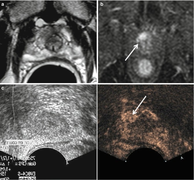
Today, urologists like Dr. Brandt are starting to use MRI as a tool to better see the cancer. So after your PSA and rectal exam, if something is detected, your doctor may send you for an MRI. If a suspicious area is identified, knowing exactly where it is can help target the next biopsy. The images from the MRI are actually used during the ultrasound biopsy as a sort of a map. One way this is done uses Prostate MRI and ultrasound together called TRUS fusion, this new technique fuses the two types of technology. Dr. Walker explains how it works:
With the TRUS fusion technique, the urologist can take an MRI image and overlay it on to their TRUS ultrasound system. The fusion technique couples real-time ultrasound with images from an MRI to help target the areas within the prostate that are suspicious to the radiologist. Dr. Brandt explains the steps:
In his office, Dr. Brandt tells his patients with an elevated PSA or an abnormal exam, If we want to get the best, most accurate biopsy that we can these days, well use an MRI ahead of doing an ultrasound-guided biopsy to accurately target any areas that are suspicious.
Where To Find The Ultrasound Equipment You Need

Is your practice ready to provide cancer screenings for men? Testicular ultrasounds, prostate ultrasounds and prostate biopsies are universally agreed upon as advanced diagnostic tools. If you want to learn even more about these ultrasound tools, . Be prepared for mens health screenings this month and find the ultrasound equipment thats right for your practice!
Looking for testicular or prostate ultrasound equipment? Contact MedCorp LLC by emailing or by calling 1-855-456-5372.
Also Check: Mri Prostate With And Without Contrast
New Ultrasound Method As Effective As Mri In Diagnosing Prostate Cancer
Researchers in the UK have demonstrated a new kind of ultrasound scan can diagnose prostate cancer with accuracy equal to costly and time-consuming magnetic resonance imaging . The findings offer clinicians an easier way to quickly test patients for clinically-significant prostate cancers.
Prostate cancer is the most common cancer to appear in men. It is a slow-growing cancer and it’s been estimated that one in five men will die with some form of asymptomatic cancer in their prostate.
Diagnosing prostate cancer can be difficult. Generally the first diagnostic method is a digital rectal examination to physically identify any prostate abnormalities. Another tool doctors have available is a blood test looking at levels of PSA a biomarker that can identity the presence of prostate cancer.
The problem with a PSA blood test is that it can often pick up signals of prostate cancer at very early stages. This leads to unnecessary treatment and anxiety in patients.
MRI scans are the most effective current tool to assess the clinical significance of prostate cancer. These scans can identify whether a cancer needs urgent clinical treatment or should be left alone for the time being.
The new method developed to investigate prostate cancer is called multiparametric ultrasound . The test uses a probe inserted into the rectum to image the prostate through a variety of ultrasound techniques.
What Happens After A Prostate Ultrasound
Once the test is done, you can take off the gown and put your clothes back on. Your rectum may feel tender for a few days, but you wont need to follow any specific aftercare instructions. Your doctor may prescribe an antibiotic to prevent infection.
In some cases, your doctor or technician may ask you to wait in the facility until your results are available. Youll usually need to wait a few days for a radiologist to look at the images and diagnose any conditions, however. Depending on where the test was done, you may wait up to two weeks for results.
Your doctor will schedule a follow-up appointment to discuss your test results. If you have any abnormalities or conditions that are visible on the images, your doctor will point out these areas. Excess tissue, prostate enlargement, or cancerous tumors will appear on the ultrasound images as bright white areas that represent the dense tissue.
Dont Miss: Does Aspirin Help Enlarged Prostate
Read Also: Is Prostate And Pancreatic Cancer The Same
Combination Biopsy Vs Mpmri
Rouviere et al report similar detection rates for standard systematic and mpMRI-targeted biopsy , but higher efficacy if the techniques are combined.89 Using both techniques in conjunction detected an additional 5.2% of csPCa over targeted biopsy alone and 7.6% over systematic biopsy alone.89 Ahdoot et al report combined biopsy led to 10% higher PCa diagnosis rate than either systematic or targeted biopsy alone and detected higher grade cancer than previously identified in 21.8% of the patients.90 A study by Oderda et al found using a combined biopsy technique increased overall cancer detection rate by 15% and the detection of csPCa by 12% over targeted biopsy alone.91 Research by Elkhoury et al further support these data, reporting a 23% increase in cancer detection over targeted biopsy alone and a 10% increase over systematic alone when using a combined technique.92 Fourcade et al report similar findings, suggesting combined biopsy increases PCa detection rate by 33% and 11.5% over targeted biopsy and systematic biopsy, respectively.93
Biopsy During Surgery To Treat Prostate Cancer
If there is more than a very small chance that the cancer might have spread , the surgeon may remove lymph nodes in the pelvis during the same operation as the removal of the prostate, which is known as a radical prostatectomy .
The lymph nodes and the prostate are then sent to the lab to be looked at. The lab results are usually available several days after surgery.
You May Like: Prostate Cancer Symptoms Blood Test
Our Study Is The First To Show That A Special Type Of Ultrasound Scan Can Be Used As A Potential Test To Detect Clinically Significant Cases Of Prostate Cancer The Can Detect Most Cases Of Prostate Cancer With Good Accuracy Although Mri Scans Are Slightly Better
Washington, October 29: According to new research, an ultrasound scan can be used to detect prostate cancer. Researchers at Imperial College London, University College London and Imperial College Healthcare NHS Trust have found that a new type of ultrasound scan can diagnose most prostate cancer cases with good accuracy in a clinical trial involving 370 men.
The ultrasound scans missed only 4.3 per cent more clinically important prostate cancer cases — cancer that should be treated rather than monitored — compared to magnetic resonance imaging scans currently used to detect prostate cancer.
MRI scans are expensive and time-consuming. The team believes that an ultrasound scan should be used as a first test in a community healthcare setting and in low and middle-income countries which do not have easy access to high-quality MRI scans. They say it could be used in combination with current MRI scans to maximise cancer detection. The study is published in Lancet Oncology. World Stroke Day 2022 Date and Theme: Know History, Significance and Ways To Observe This Day Started by World Stroke Organization To Raise Awareness About the Ailment.
Professor Hashim Ahmed, lead author of the study and Chair of Urology at Imperial College London, said: “Prostate cancer is the most commonly diagnosed cancer in the UK. One in six men will be diagnosed with the disease in their lifetimes and that figure is expected to rise.
Research: Ultrasound Scan Can Detect Prostate Cancer
Researchers at Imperial College London, University College London and Imperial College Healthcare NHS Trust have found that a new type of ultrasound scan can diagnose most prostate cancer cases with good accuracy in a clinical trial involving 370 men. Read on for details:
Washington : According to new research, an ultrasound scan can be used to detect prostate cancer.
Researchers at Imperial College London, University College London and Imperial College Healthcare NHS Trust have found that a new type of ultrasound scan can diagnose most prostate cancer cases with good accuracy in a clinical trial involving 370 men.
The ultrasound scans missed only 4.3 per cent more clinically important prostate cancer cases — cancer that should be treated rather than monitored — compared to magnetic resonance imaging scans currently used to detect prostate cancer.
MRI scans are expensive and time-consuming. The team believes that an ultrasound scan should be used as a first test in a community healthcare setting and in low and middle-income countries which do not have easy access to high-quality MRI scans. They say it could be used in combination with current MRI scans to maximise cancer detection. The study is published in Lancet Oncology.
As cancer waiting lists build as a result of the COVID-19 pandemic, there is a real need to find more efficient and cheaper tests to diagnose prostate cancer.
Recommended Reading: What Percent Of Prostate Cancers Are Aggressive
Researchers Have Found That A New Type Of Ultrasound Scan Can Detect Most Prostate Cancer Cases With Good Accuracy
According to new research, an ultrasound scan can be used to detect prostate cancer. Researchers at Imperial College London, University College London and Imperial College Healthcare NHS Trust have found that a new type of ultrasound scan can diagnose most prostate cancer cases with good accuracy in a clinical trial involving 370 men.
The ultrasound scans missed only 4.3 per cent more clinically important prostate cancer cases — cancer that should be treated rather than monitored — compared to magnetic resonance imaging scans currently used to detect prostate cancer. MRI scans are expensive and time-consuming. The team believes that an ultrasound scan should be used as a first test in a community healthcare setting and in low and middle-income countries which do not have easy access to high-quality MRI scans. They say it could be used in combination with current MRI scans to maximise cancer detection. The study is published in Lancet Oncology.
“Our study is the first to show that a special type of ultrasound scan can be used as a potential test to detect clinically significant cases of prostate cancer. The can detect most cases of prostate cancer with good accuracy, although MRI scans are slightly better. “We believe that this test can be used in low and middle-income settings where access to expensive MRI equipment is difficult and cases of prostate cancer are growing.”
New Ultrasound Scan Technology

Finding new ways to diagnose prostate cancer that are cost-effective, and time-saving is essential. MRI scans are often used in the diagnosis process however, this method is expensive. The research team believe that an ultrasound scan should be used as the first test in a community healthcare setting. It also has potential for use in low-and-middle-income countries which do not have easy access to high-quality MRI scans.
Professor Hashim Ahmed, lead author of the study and Chair of Urology at Imperial College London, said: Prostate cancer is the most commonly diagnosed cancer in the UK. One in six men will be diagnosed with the disease in their lifetimes and that figure is expected to rise.
Our study is the first to show that a special type of ultrasound scan can be used as a potential test to detect clinically significant cases of prostate cancer. They can detect most cases of prostate cancer with good accuracy, although MRI scans are slightly better.
The new study analysed the use of a different kind of ultrasound scan technology called multiparametric ultrasound , which uses soundwaves to look at the prostate. The test involves a probe called a transducer to make the images of the prostate and is placed in the rectum. Following this, it sends waves that bounce off the organs and other structures. These are made into pictures of the organs.
Read Also: Does Super Beta Prostate Really Work
How Is Prostate Cancer Diagnosed
A biopsy is when a small piece of tissue is removed from the prostate and looked at under a microscope.
A biopsy is a procedure that can be used to diagnose prostate cancer. A biopsy is when a small piece of tissue is removed from the prostate and looked at under a microscope to see if there are cancer cells.
A Gleason score is determined when the biopsy tissue is looked at under the microscope. If there is a cancer, the score indicates how likely it is to spread. The score ranges from 2 to 10. The lower the score, the less likely it is that the cancer will spread.
A biopsy is the main tool for diagnosing prostate cancer, but a doctor can use other tools to help make sure the biopsy is made in the right place. For example, doctors may use transrectal ultrasound or magnetic resonance imaging to help guide the biopsy. With transrectal ultrasound, a probe the size of a finger is inserted into the rectum and high-energy sound waves are bounced off the prostate to create a picture of the prostate called a sonogram. MRI uses magnets and radio waves to produce images on a computer. MRI does not use any radiation.
Most Common Prostate Exams
The most common prostate screening exams are 1.) a blood test that checks your PSA level and 2.) a rectal exam. Unless either of these point to a problem, or youre having symptoms like difficulty going to the bathroom, the PSA and the rectal exam are likely all youll ever have. But, if you have an abnormal exam or cancer is detected, you can expect a biopsy. The industry standard is a transrectal ultrasound biopsy performed in your urologists office. Dr. Brandt says the TRUS biopsy does have shortfalls. In some patients, a biopsy is negative but their PSA level is still rising. It may mean the ultrasound didnt see the cancer. Dr. Sidney Walker, a body radiologist at RAYUS, says a TRUS biopsy isnt always able to identify the prostate cancer because of where the cancer lesion is located. Theres actually a region of your prostate that is hard to see with ultrasound says Dr. Brandt:
An ultrasound isnt necessarily all that sensitive for picking up areas or specific parts of the prostate or zones of the prostate which may harbor the cancer. That is, we can look with an ultrasound but we may not actually see the cancer.
Recommended Reading: What Foods Help Prostate Health
