Two Agents Approved For Psma
On May 26, FDA approved piflufolastat F 18 for use in a type of imaging procedure called PSMA PET in people with prostate cancer. The approval covers the use of piflufolastat F 18 in patients suspected of having metastatic prostate cancer or recurrent prostate cancer . Last year, the agency approved another imaging agent for PSMA PET, Ga 68 PSMA-11, for the same uses, but its use is largely limited to the two institutions where it is made.
In a statement, Lantheus, which manufactures piflufolastat F 18, said the imaging agent will be immediately available in parts of the mid-Atlantic and southern regions with broad availability across the U.S. anticipated by year end.
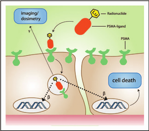
PSMA is often overproduced by prostate cancer cells but is generally not produced by most normal cells, making it an excellent target for both PET imaging and targeted systemic radiation therapy like 177Lu-PSMA-617, Dr. Morris said.
How Does The Psma Pet Work Technical Aspects
This imaging technique is based on a small-molecule PSMA binding chemical attached to a radioactive reporter. The PET tracer is consequently composed of the PSMA-binding and the radioactive reporter . This tracer, resulted by means of the conjugation of the aforementioned elements, is introduced in the patients circulation. The main property of this conjugate is that it accumulates at prostate cancer sites, highlighting the areas that are affected by the tumor. An imaging camera makes these areas visible and records the places where the PET tracer accumulated. The unbound conjugate is rapidly cleared from circulation.
This FDA approval is based on intensive studies made at the Universities of California, San Francisco, and Los Angeles. These studies, funded by the Prostate Cancer Foundation, demonstrated the level of sensitivity this imaging technique is having. All the clinical trials have proved that it can be used in order to detect recurrent prostate cancer in men with high PSA levels after prostate cancer treatment and even for detecting metastases in men first diagnosed with high-risk prostate cancer.
Who Can Benefit From This New Diagnosis Method
Patients who are first diagnosed with high-risk prostate cancer can undergo a PSMA PET scan in order to have a clearer view of the tumor expansion. Also, patients who have had prostate cancer treatment and have rising PSA can benefit from the advantages of the new PSMA PET to localize the new tumor. In both cases, imaging techniques are extremely important. Why? First of all, doctors can know that a patient is having cancer that is growing by means of the PSA test. That is a blood test that checks for Prostate Specific Antigens in the blood. If, after treatment, the PSA is rising, doctors know that the patient is dealing with recurrent cancer. The problem is that they cannot clearly identify where it is located. Not knowing where the cancer is, doctors cannot treat it. Is surgery an option or external beam radiation? Exact localization by means of PSMA PET empowers doctors with the ability to design a specific care plan.
Studies are being done in order to determine how this new imaging technique will reshape the manner in which prostate cancer treatments are being prescribed and, furthermore, patients survival rates.
Read Also: Can Losing Weight Shrink Prostate
How To Get A Psma Pet Scan
- PSMA PET scans are offered at UCSF Radiology China Basin location in the San Francisco Bay area.
- For UCSF patients, please reach out to Radiology Scheduling directly to schedule your PSMA scan.
- For non-UCSF facilities referring patients that are new to UCSF, please fax the following to 353-7299 for patient registration to be completed prior to scheduling:
- Patient Exam Order
- Insurance Information and Authorization
How Do Psma Pet Scans Work And What Are Their Benefits

Weve been using PET imaging to detect cancers for decades. Until recently, however, we havent been able to image prostate cancer well because older PET radiotracers these are radioactive molecules that stick to cancer cells and can be seen on PET scans do not routinely bind to prostate cancer. However, a new radiotracer, piflufolastat F-18 , does bind to the PSMA on prostate cancer cells. Doctors can look at a PET scan and see if piflufolastat lights up prostate cancer cells anywhere in the body. Today, PSMA PET is the single best method we have to precisely visualize prostate cancer in the body. Knowing the anatomic location of prostate cancer helps us make better treatment decisions and monitor a patients response to therapy.
Also Check: Natural Herbs To Shrink Prostate
Also Check: Does Prostate Massage Help Bph
What Does This Mean To You
While reading this article, you may have wondered how this research affects you. Practically, it means that now it has become much easier to precisely diagnose high-risk prostate cancer tumors, localize them and, eventually, treat them accordingly. But it all starts with patients awareness of the disease, which can give no symptoms while in the early stages. That being said, do not postpone a urologist appointment. Early screenings can be life-saving and health-caring should always be a priority. Would you like to know more about what a screening procedure involves? This article provides useful insights on prostate cancer screening and diagnosis. Take informed steps today for better health tomorrow!
Axumin Pet Scanning For Prostate Cancer Care
Axumin is an FDA-approved agent used for Axumin PET scans for prostate cancer. Axumin is often able to image and restage recurrent prostate cancer better than any other conventional imaging techniques. Biochemical recurrence, typically suspected with rising PSA levels, is the standard in monitoring patients for suspected recurrent prostate cancer. Traditional imaging techniques are often limited in that they may detect a small lymph node or suspicious finding, but cannot further functionally characterize the molecular activity to determine the level of suspicion. The introduction of Axumin PET scanning has been a breakthrough, allowing physicians the ability to accurately locate and restage prostate cancer with precision, especially in the setting of suspiciously rising PSA levels.
How Do Axumin PET Scans Work?
An Axumin PET uses a radioactive tracer, given as an injection, that is linked to an amino acid which is absorbed by prostate cancer at a much more rapid rate than normal cells. The rapid uptake of Axumin by prostate cancer cells is then imaged by the advanced technology within the PET scan equipment. The PET scan images are then reviewed in order to determine if there has been any spread to other areas in the body.
Recommended Reading: Do Doctors Milk Your Prostate
What Is The Psma Pet Scan For Prostate Cancer
The PSMA PET scan is a test that can help your doctor learn if and where prostate cancer has spread outside your prostate gland, including to your lymph nodes, other organs, or bones.
PET scans are a type of imaging test that use special dye with radioactive tracers to make cancer cells show up more clearly.
The PSMA PET scan uses radioactive tracers that bind to prostate-specific membrane antigen . This is a protein thats found in high levels on the surface of prostate cancer cells.
The Food and Drug Administration has recently approved the following PSMA-targeted tracers:
Researchers are studying other PSMA-targeted tracers, which might be approved in the future.
What Is The New Psma Pet
The abbreviation PSMA comes from Prostate-Specific Membrane Antigen. This Membrane Antigen is a protein that is found in larger amounts on prostate cancer cells. Doctors are able to visualize these proteins by means of a PET scanner. To date, there are also other types of imaging diagnosis techniques for prostate cancer, such as CT, MRI, or bone scans. However, the PSMA PET is known as the most sensitive way to find prostate cancer in the world right now.
The PSMA image technique is a method used to identify the presence of small tumor cells. It can be used in order to distinguish between the nature of the tissue, whether there are tumor signs or not. What is more, it can detect metastasis much earlier, when they are smaller. Doctors can find almost any prostate cancer if its bigger than a pea bean in size. By means of this technique, doctors can see very specifically where the sites of prostate cancer are. Is it limited to the prostate? Is it in the bones or is it in the lymph nodes? This gives doctors the ability to design treatment approaches for that specific patient depending on his specific circumstances.
This, in turn, results in treatments with higher success rates.
Don’t Miss: When Do Prostate Exams Start
The Future Of Psma Pet
This is a solid study and reflects the real-world experience with PSMA PET-CT in other countries, Dr. Pomper said. Because there are several PSMA-targeted tracers, a next step will be to have them approved for use in the United States outside of clinical trials, he added.
He predicted that, eventually, the different PSMA tracers will be tested head to head.
The Australian trial adds to a growing body of research on improving the detection of metastatic tumors in men with prostate cancer. One imaging agent, fluciclovine F18 which targets prostate cancer cells in a different way than PSMA-targeted tracersis already approved in the United States for use in men with previously treated prostate cancer that appears to be progressing .
PSMA PET-CT is also being studied in this group of men, Dr. Shankar said. One small clinical trial that directly compared PSMA PET-CT with fluciclovine F18 PET-CT showed that the PSMA-targeted scan found more metastatic tumors, regardless of their location. NCI is funding a similar but larger clinical trial.
Dr. Pomper noted that PSMA also is found at relatively high levels in the vasculature of a number of other cancersincluding kidney, thyroid, and breastso hes hopeful that PSMA PET-CT might be useful beyond prostate cancer.
Urologists and radiation oncologists in many places are already ordering this scan as the standard of care, he said.
Things To Know About Psma
If you have your ear to the ground regarding all things prostate cancer you may have heard some buzz about PSMA-PET and want to know more. On the other hand, maybe youve never heard of PSMA-PET before in your life. Weve got you covered either way, here are the 5 things you need to know about the newly FDA approved PSMA-PET scan.
Also Check: Risk Factors For Prostate Cancer
How Psma Fuels Prostate Cancer
Now a team of researchers led by MSK radiologist and molecular imaging specialist has discovered that PSMA plays a role in prostate cancers development. They report in the Journal of Experimental MedicinethatPSMA indirectly activates a cancer-causing pathway that involves the PI3K protein kinase.
Everybody knew that PSMA was related to more-aggressive prostate cancer, but nobody was able to figure out its biological role, Dr. Grimm says. Knowing how it helps trigger PI3K has important implications for treating men with prostate cancer.
In cell and animal experiments, as well as in tumor samples from patients, Dr. Grimms team found that PSMA causes the release of the amino acid glutamate, which acts as a second messenger to bind to a receptor on the surface of prostate cells. This binding transmits a signal to the cell to help activate the PI3K pathway. Dr. Grimms team showed that targeting PSMA with drugs slowed PI3K signaling, as well as the growth and spread of PSMA-expressing tumors, and, in the animal subjects, allowed them to live longer.
Dr. Grimm explains that PSMA inhibitors could potentially be used in combination with the existing prostate cancer drugs called androgen inhibitors, which block hormones that fuel cancer growth.
How Do I Prepare For A Psma Pet Scan

Preparation
You May Like: How To Reduce Prostate Enlargement Naturally
Greater Accuracy And Changing Treatment
Approximately 300 men were enrolled in the Australian trial, all with newly diagnosed localized prostate cancer , and all were considered to have high-risk disease. For all men in the trial, the planned treatment was either surgery or radiation therapy to the prostate only.
Half the men were randomly assigned to initially undergo a CT and bone scan, and the other half to PSMA PET-CT.
Based on the imaging, PSMA PET-CT was 27% more accurate than the standard approach at detecting any metastases . Accuracy was determined by combining the scans sensitivity and specificity, measures that show a tests ability to correctly identify when disease is present and not present.
PSMA PET-CT was more accurate for both metastases found in lymph nodes in the pelvis and in more distant parts of the body, including bone. Radiation exposure was also substantially lower with PSMA PET-CT than with the conventional approach.
The trial investigators also tracked how imaging results influenced clinicians treatment choices. Based on imaging findings, the initial treatment plan was changed for 15% of men who underwent conventional imaging compared with 28% of men who underwent PSMA PET-CT.
Another key finding, Dr. Hofman noted, was that PSMA PET-CT was much less likely to produce inconclusive, or equivocal, results .
Thats important, he continued, because if you have a scan with equivocal findings, it often leads to more scans or biopsies or other tests.
Has The Radiotracer Used In Psma Pet Scans Been Approved By The Fda
The radiotracer piflufolastat, also known as Pylarify, is safe and FDA-approved. Side effects are rare and temporary and may include headache and a change in taste. Piflufolastat is a radionuclide dye, so it contains a small amount of radiation. The amount is similar to the exposure you receive during a CT scan and the tracers radioactive element is entirely gone from your body within a few days.
Read Also: Where In The Body Is The Prostate
Psma And Clinical Management
The results of a retrospective, real-world, single-institution study showed that PSMA PET/CT after conventional imaging may lead to management changes in up to 60% of patients.15,16
Eur J Nucl Med Mol Imaging
Furthermore, based on preclinical data, PSMA has been shown to affect several key oncogenic pathways* and is being evaluated as a relevant therapeutic target.18-23
Cell proliferation and survival18-21
Angiogenesis22,23
Many of these outcomes are driven by the role of PSMA in the PI3K/Akt pathway.18
J Exp Medin vitro
PSMA is a diagnostic biomarker and potential therapeutic target, enabling a phenotypic precision medicine approach to help guide patient selection for therapy in advanced prostate cancer.6,10,15,16,24,25
What Are The Benefits Of Psma Pet Treatment At Ucsf
- FDA approved imaging technique for prostate cancer.
- The PSMA PET scan can identifiy cancer that is often missed by current standard-of-care imaging techniques.
- The PSMA tracer can also be used in conjunction with CT or MRI scans.
- UCSF is only one of two medical centers in the U.S. that offers the FDA approved PSMA PET.
- PSMA PET is more effective and precise for localizing mestatic prostate cancer.
- UCSF researchers, along with colleagues at UCLA, studied PSMA PET for several years to better precisely locate prostate cancer.
- PSMA PET works using a radioactive tracer, called 68Ga-PSMA-11, which is manufactured on site at UCSF.
You May Like: Can Prostate Cancer Go Away
A Different Way To Detect Metastases
Most men diagnosed with prostate cancer have localized disease, meaning the cancer appears to be confined to the prostate gland. However, certain factors have been linked to a higher risk of the cancer eventually spreading .
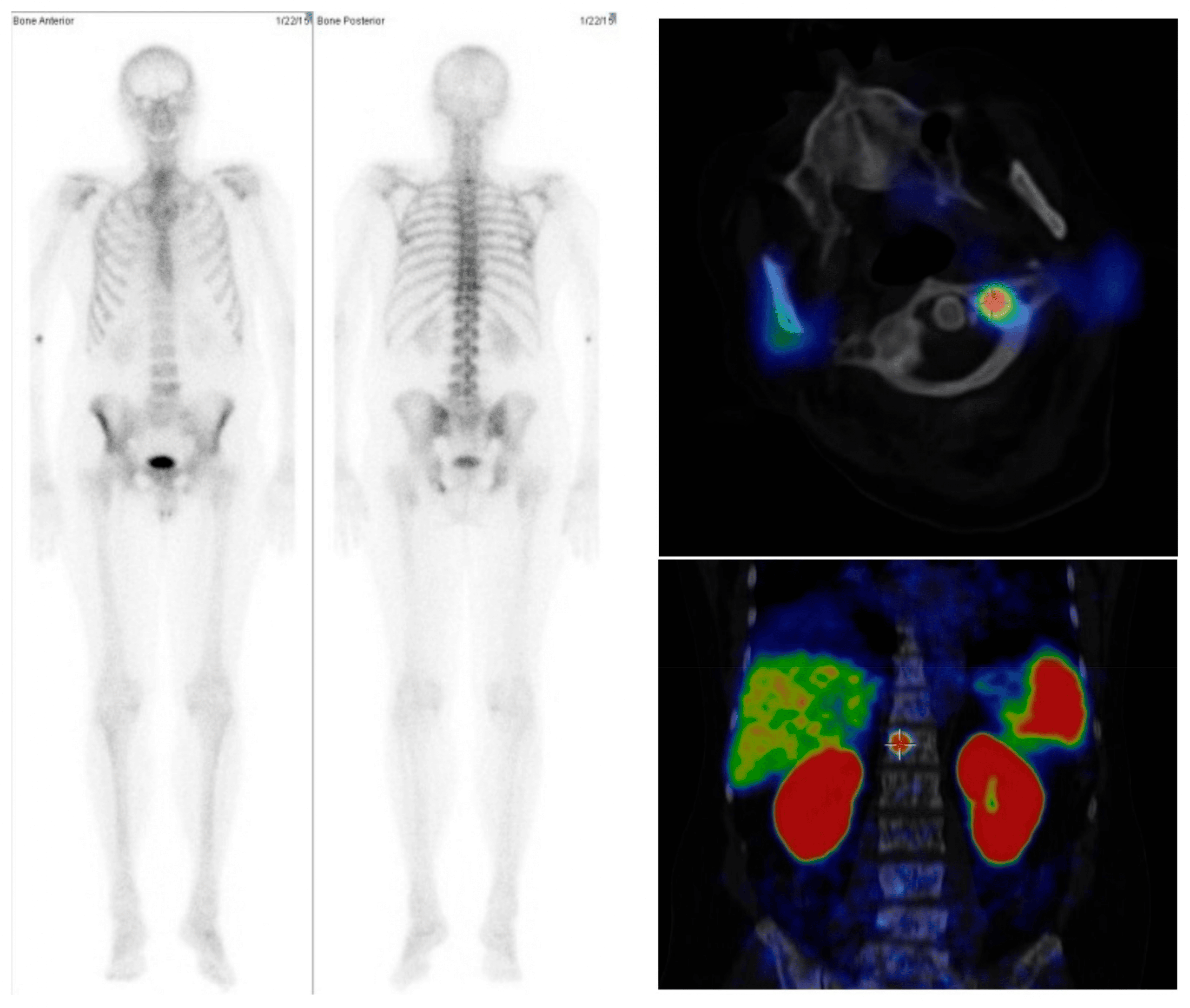
Currently, in the United States and many other countries, most men diagnosed with high-risk localized prostate cancer undergo additional testing to see if there is evidence of metastatic cancer. For many years, that has been done with a conventional CT scan and a bone scan , the latter because prostate cancer often spreads to the bones.
But both imaging technologies have limitations. Neither is particularly good at finding individual prostate cancer cells, and thus can miss very small tumors. And bone scans can detect bone damage or abnormalities that were caused by something other than cancer , resulting in false-positive findings that can lead to unnecessary additional testing.
So, researchers have been developing and testing other imaging agents that can find prostate cancer cells specifically in the body, Dr. Shankar explained.
As their name implies, PET-CT scans combine a CT scan with a PET scan, another type of nuclear imaging test that requires patients to receive intravenous injections of a radioactive tracer that can be detected on the scan.
How Long Does A Psma Scan Take
The PSMA PET scan usually takes about 2 hours, although timing may vary.
To conduct a PSMA PET scan, a nurse or technician will inject a special dye with a radioactive tracer into one of your veins. They will ask you to wait approximately 30 to 60 minutes to allow the dye to travel throughout your body.
Next, they will ask you to lie down on a padded exam table. They will slide the table through a PET-CT or PET-MRI scanner to create images of your body. This scan may take 30 minutes or longer to complete.
After the scan is finished, a specialist will review the images and report the results to your doctor. Your doctor will share the results with you.
Ask your doctor how long it will take to receive the results of the scan.
Don’t Miss: How To Get Rid Of Prostate Cancer
