Determining Whether Prostate Cancer Is Aggressive
If a biopsy sample is found to contain cancer, the pathologist analyzing the specimen takes a deeper look at the cancer cells to determine how aggressive the disease is likely to be.
If the cancer cells appear significantly abnormal and dissimilar from healthy cells under a microscope, the cancer is considered more aggressive and expected to advance quickly. Conversely, cancer cells that look relatively similar to healthy cells indicate that its less aggressive and may not spread as fast.
Prostate cancers are assigned a Gleason score depending on how abnormal the cells look..
Gleason score: Gleason scores range from 2 to 10, going from least to most aggressive prostate cancers.
There are different types of cancer cells in a prostate tumor, so the final Gleason score is determined by adding the scores of the two main areas of the tumor.
First, the primary part of the tumor is assigned a number between 1 and 5. Lower numbers indicate that the cells appear relatively similar to healthy cells, while higher numbers show that the cells are abnormal-looking. Then, another number between 1 and 5 is assigned to describe the second most prevalent area of the tumor.
Finally, the two numbers assigned to the different parts of the prostate tumor are added. So, if most of the tumor is given a 4, and some of the tumor is more aggressive and given a 5, the final Gleason score would be 9.
There are many biomarker tests, including:
- Oncotype DX® Genomic Prostate Score
- Prolaris
- ProMark®
What Is A Transrectal Ultrasound
The digital rectal examination and the prostate-specific antigen blood test are two important ways to detect changes in the prostate gland.
However, they cannot determine if the changes are due to prostate cancer or to a non-cancerous condition. In the event of a significantly elevated PSA test and/or abnormal DRE, a prostate needle biopsy the surgical removal of tissue for examination under a microscope must be performed in order to make a definitive diagnosis of prostate cancer. The biopsy is taken with the guidance of transrectal ultrasound.
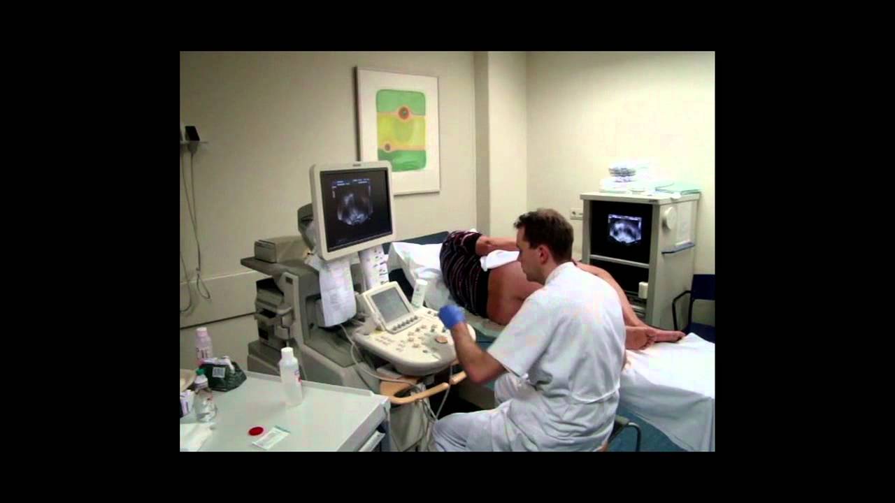
Transrectal ultrasound is a 5- to 15-minute outpatient procedure that uses sound waves to create a video image of the prostate gland. A small, lubricated probe placed into the rectum releases sound waves, which create echoes as they enter the prostate.
Prostate tumors sometimes create echoes that are different from normal prostate tissue. The echoes that bounce back are sent to a computer that translates the pattern of echoes into a picture of the prostate. While the probe may be temporarily uncomfortable, TRUS is essentially a painless procedure.
Enhanced Ultrasound Improves Detection Of Prostate Cancer
According to a recent article published in The Journal of Urology, the use of contrast enhanced color Doppler in endorectal ultrasound improves the detection of prostate cancer.
Prostate cancer is a common cancer among men in the United States. Prostate cancer is the second leading cause of cancer death in men in the United States. The prostate is a walnut-size gland that is located between the bladder and rectum and forms a component of semen. Prostate specific antigen levels , a digital rectal exam and transrectal ultrasound are common tests used to detect prostate cancer. If any suspicious mass is found through these tests, a patient must then undergo biopsies to definitively determine whether cancer exists. However, it is imperative that a physician takes a biopsy from the area in the prostate where the cancer exists to provide accurate diagnostic information. Physicians often use endorectal ultrasound to help determine where in the prostate to take a biopsy. Researchers are attempting to improve upon the accuracy of ultrasound in the guidance of placement of biopsies, including the introduction of contrast, which help physicians to discern between healthy looking tissue and possible sites of cancer.
Reference: Roy C, Buy X, Lang H, et al. Contrast enhances color Doppler endorectal sonography of the prostate: efficiency for detecting peripheral zone tumors and role for biopsy procedure.
The Journal of Urology. 2003 170:69-72.
Read Also: How Long Does It Take For Prostate Cancer To Spread To The Bones
Prostate Cancer: Advancements In Screenings
Reviewed By:
Dr. Christian Paul Pavlovich
You may know thatprostate canceris one of the most common cancer types in men. The good news is that thereare many treatment and management options, even if the cancer is caught ata later stage.
What you may not know: There are several options when it comes toprostate cancer screening. After considering multiple factors, your doctor may recommend theprostate-specific antigen test, and/or one of the newer screeningtests that are now available.
Johns Hopkins urologistChristian Pavlovich, M.D., explains what you should know.
Dont Miss: What Is Perineural Invasion In Prostate Cancer
What Are The Advantages And Disadvantages Of Hifu

What may be important for one person might not be so important for someone else. If youre thinking about having HIFU, speak to your doctor or nurse before deciding whether to have it. They can help you choose the right treatment for you. Take time to think about whether you want to have HIFU.
Advantages
- HIFU doesnt involve any cuts to the skin or needles, apart from a needle in your hand to give you a general anaesthetic.
- Focal HIFU can treat small areas of cancer while causing little damage to nearby tissue, nerves and muscles.
- You only need a short hospital stay you can usually go home on the same day as your treatment.
- Recovery is usually quick and most men return to their normal activities within two weeks.
- HIFU is less likely than surgery to cause erection or urinary problems.
- You may be able to have HIFU if your cancer has come back after radiotherapy.
- You may be able to have HIFU again if your cancer comes back after your first HIFU treatment. This isnt the case with all treatments.
- You may also be able to have other treatments after HIFU if your cancer comes back, such as surgery or radiotherapy.
Disadvantages
- In the UK, HIFU is only available in specialist centres or as part of a clinical trial. You may need to travel a long way to your nearest treatment centre.
- Compared with other treatments, we dont know how well it works in the longer term .
- As with other treatments, you may get side effects, such as erection and urinary problems.
Read Also: How Long Should You Take Lupron For Prostate Cancer
Seeing The Cancer With Mri
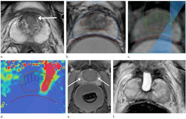
Today, urologists like Dr. Brandt are starting to use MRI as a tool to better see the cancer. So after your PSA and rectal exam, if something is detected, your doctor may send you for an MRI. If a suspicious area is identified, knowing exactly where it is can help target the next biopsy. The images from the MRI are actually used during the ultrasound biopsy as a sort of a map. One way this is done uses Prostate MRI and ultrasound together called TRUS fusion, this new technique fuses the two types of technology. Dr. Walker explains how it works:
With the TRUS fusion technique, the urologist can take an MRI image and overlay it on to their TRUS ultrasound system. The fusion technique couples real-time ultrasound with images from an MRI to help target the areas within the prostate that are suspicious to the radiologist. Dr. Brandt explains the steps:
In his office, Dr. Brandt tells his patients with an elevated PSA or an abnormal exam, If we want to get the best, most accurate biopsy that we can these days, well use an MRI ahead of doing an ultrasound-guided biopsy to accurately target any areas that are suspicious.
Preparing For Your Pet
For most PET-CT scans, you need to stop eating about 4 to 6 hours beforehand. You can usually drink water during this time. You might have instructions not to do any strenuous exercise for 24 hours before the scan.
Some people feel claustrophobic when theyre having a scan. Contact the department staff before your test if youre likely to feel like this. They can take extra care to make sure youre comfortable and that you understand whats going on.
Your doctor can arrange to give you medicine to help you relax, if needed.
Also Check: How Effective Is Chemotherapy For Prostate Cancer
Accurate Diagnosis Of Prostate Cancer With Ultrasound
- Date:
- Eindhoven University of Technology
- Summary:
- Prostate cancer is the most common type of cancer among men, but its diagnosis has up to now been inaccurate and unpleasant. Researchers have now developed an imaging technology that can accurately identify tumors. The technology is based on ultrasound, and also has the potential to assess how aggressive tumors are. This can lead to better and more appropriate treatment, and to cost savings in health care.
Prostate cancer is the most common type of cancer among men, but its diagnosis has up to now been inaccurate and unpleasant. Researchers at Eindhoven University of Technology in the Netherlands, in cooperation with AMC Amsterdam, have developed an imaging technology that can accurately identify tumors. The technology is based on ultrasound, and also has the potential to assess how aggressive tumors are. This can lead to better and more appropriate treatment, and to cost savings in health care.
About 11% of men who die of cancer in the western world do so as a result of prostate cancer. Each year 200,000 men are diagnosed with the disease in the US alone. But diagnosis is still rudimentary. After determining the PSA level in the blood-, biopsies are performed to see if there are tumors in the prostate. However the PSA level is not a very good indicator: two-thirds of all biopsies turn out to have been unnecessary.
Biopsies are not targeted
Aggressive
No biopsies
Story Source:
What Are The Limitations Of Prostate Ultrasound Imaging
Men who have had the tail end of their bowel removed during prior surgery are not good candidates for ultrasound of the prostate gland because this type of ultrasound typically requires placing a probe into the rectum. However, the radiologist may attempt to examine the prostate gland by placing a regular ultrasound imaging probe on the perineal skin of the patient, between the legs and behind the scrotum of the patient. Sometimes the gland can be examined by ultrasound this way, but the images may not be as detailed as with the transrectal probe. An MRI of the pelvis may be obtained as an alternative imaging test, because it may be obtained with an external receiver coil.
Also Check: Do Females Have Prostate Cancer
How Do I Prepare For A Prostate Ultrasound
Some possible instructions that your doctor might give you before the test include:
- Dont eat for a few hours before the test.
- Take a laxative or enema to help clear out your intestines a few hours before the test.
- Stop taking any medications that can thin your blood, such as nonsteroidal anti-inflammatory drugs or aspirin, about a week before the procedure. This is usually recommended if your doctor plans to take a biopsy of your prostate.
- Dont wear any jewelry or tight clothes to the clinic on the day of the procedure.
- Take any medications recommended to help you relax during the procedure. Your doctor may recommend a sedative, such as lorazepam .
- Make sure someones available to take you home in case your doctor gives you a sedative.
What Are Some Common Uses Of The Procedure
A transrectal ultrasound of the prostate gland is performed to:
- detect disorders within the prostate.
- determine whether the prostate is enlarged, also known as benign prostatic hyperplasia , with measurements acquired as needed for any treatment planning.
- detect an abnormal growth within the prostate.
- help diagnose the cause of a man’s infertility.
A transrectal ultrasound of the prostate gland is typically used to help diagnose symptoms such as:
- a nodule felt by a physician during a routine physical exam or prostate cancer screening exam.
- an elevated blood test result.
- difficulty urinating.
Because ultrasound provides real-time images, it also can be used to guide procedures such as needle biopsies, in which a needle is used to sample cells from an abnormal area in the prostate gland for later laboratory testing.
You May Like: Prostatitis Viagra
Why Is A Pet/ct Scan Better Than A Ct Scan
A PET/CT scan, meanwhile, does show if tissue is cancerous or not, and how active it is. This means you wont receive a false negative. Plus, you will avoid continuing treatment if its not actually needed. Weve had clients whod been told that they were cancer-free, based on a CT scan. But when they got a PET/CT scan, it showed a cancerous tumour that would have otherwise gone untreated.
Simply put, you cannot rely on a CT scan. See our PET/CT vs CT Comparison to learn more about how and why CT scans can fail cancer patients.
Recommended Reading: Prostate Biopsy Results Timeline
What Happens During A Prostate/rectal Ultrasound

You may have a prostate/rectal ultrasound done as an outpatient or during ahospital stay. The way the test is done may vary depending on yourcondition and your healthcare providers practices.
Generally, a prostate/rectal ultrasound follows this process:
You will need to remove any clothing, jewelry, or other objects that may get in the way of the procedure.
If asked to remove clothing, you will be given a gown to wear.
You will lie on an exam table on your left side with your knees bent up to your chest.
The healthcare provider may do a digital rectal exam before the ultrasound.
The provider puts a clear gel on the transducer and puts the probe into the rectum. You may feel a fullness of the rectum at this time.
The provider will turn the transducer slightly several times to see different parts of the prostate gland and other structures.
If blood flow is being looked at, you may hear a whoosh, whoosh sound when the Doppler probe is used.
Once the test is done, the provider will wipe off the gel.
A prostate/rectal ultrasound may be uncomfortable and you will need toremain still during the test. The gel will also feel cool and wet. Thetechnologist will use all possible comfort measures and do the scan asquickly as possible to minimize any discomfort.
Also Check: Is Zinc Good For Prostate
What Is The Strength Of The Mri Scanner
The field strength of the MRI magnet is measured in teslas, and systems can range between .5T and 3T. A 3T scanner will provide the radiologist with the highest quality images in the case of the prostate gland which is a very small organ. Images from a scanner with a weaker field strength could potentially lead to an inaccurate diagnosis. We give a lot of second opinions of scans that have been performed elsewhere, says Dr. Shaish. Often the quality isnt high enough, and we cant provide the diagnosis that the patient needs.
When To Consider A Prostate Mri
Studies indicate that MRI may be helpful in the following situations. The best images are obtained when using an endorectal coil.
- You have a PSA that continues to increase, but an ultrasound-guided prostate biopsy does not reveal cancer an MRI may be able to better pinpoint a suspicious area for a more targeted biopsy and increase the likelihood of finding cancer if it is there.
- Different elements of your diagnostic workup are in conflict an MRI can better determine size of the tumor and whether it has extended beyond the capsule.
- For large palpable tumors, MRI can rule out cancer that extends beyond the prostate itself.
- If your PSA rises following prostate cancer treatment, MRI can be used to identify any cancerous tissue in the periprostatic bed , which indicates a local recurrence.
- MRI may provide better guidance about where to target radiation therapy.
Don’t Miss: Does Enlarged Prostate Affect Ejaculation
Early Detection Saves Lives
Prostate cancer is the most common cancer affecting Australian men .
Prostate cancer is the growth of abnormal cells in the prostate gland. This gland is only found in males and is about the size of a walnut.
The causes of prostate cancer are not understood and there is currently no clear prevention strategy.
Preparing For The Scan
You usually have this scan in the hospital x-ray department.
You need to make sure your bowel is empty when you go for your appointment. You might need to have an enema to empty your bowel. An enema is a liquid that you put into your back passage .
Or you might have a liquid medicine to swallow the day before. You need to stay close to a toilet for a few hours after taking the medicine.
Check your appointment letter to find out how to prepare for your scan.
Also Check: Prostate Cancer Chemo Side Effects
Who Interprets The Results And How Do I Get Them
A radiologist, a doctor trained to supervise and interpret radiology exams, will analyze the images. The radiologist will send a signed report to the doctor who requested the exam. Your doctor will then share the results with you. In some cases, the radiologist may discuss results with you after the exam.
Follow-up exams may be needed. If so, your doctor will explain why. Sometimes a follow-up exam is done because a potential abnormality needs further evaluation with additional views or a special imaging technique. A follow-up exam may also be done to see if there has been any change in an abnormality over time. Follow-up exams are sometimes the best way to see if treatment is working or if an abnormality is stable or has changed.
