Breaking News: Fda Approves A Highly Sensitive Prostate Cancer Imaging Agent
What this means for patients: Today, the FDA approved another highly sensitive imaging compound specifically for prostate cancer called 18F-DCFPyL. This is the second such approval in less than six months in December, the FDA approved 68Ga-PSMA-11 PET. These pioneering new scanning tools will revolutionize prostate cancer detection. Both imaging agents are used to light up PET scans to help doctors find smaller tumors earlier.
Both compounds are part of a new type of scanning technology called PSMA PET imaging. 68Ga-PSMA-11 and18F-DCFPyL are radioactive tracer molecules designed to bind to PSMA that doctors can use to light up PET scans for regions that contain cancer. PSMA is a protein found on the surface of prostate cancer cells. This new technology is more sensitive than conventional imaging in finding areas of prostate cancer in the body.
Having more complete and accurate information about where cancer is located can help doctors make better treatment plans. Finding metastases earlier, when they are much smaller, will have a significant impact for patients.
PyL PET imaging is approved for two types of patients with prostate cancer: 1) those with suspected metastasis who are candidates for initial definitive therapy and 2) those with suspected recurrence based on elevated PSA level .
Read more about PyL here and about the amazing science behind PSMA here
Prostate Cancer In Australian Men
Written by Dr Eamonn McAteer & Dr David Homewood, Radiologists and Partners, South Coast Radiology.
Prostate cancer has become the most significant major malignancy of Australian men and is responsible for 3,300 deaths annually. Around 20,000 new cases are diagnosed every year, yet diagnosing this condition still presents a challenge.
What Happens During The Psma Pet Scan
The day before your scheduled PSMA PET/CT, a nurse will call you to ask you about your medications and remind you of your appointment time. The exam doesnt require fasting. And patients are encouraged to drink fluids before and after their exam.
After arriving for your scan, you will be asked your height and weight. Youll also be asked to remove metal objects such as jewelry and belt buckles. A nurse will place an IV in your arm. And a technologist will use the IV to inject radioactive PSMA. The IV will be removed after the injection. Youll wait in a comfortable room for an hour while the PSMA circulates and attaches to prostate cancer cells.
Also Check: How To Get To A Man’s Prostate
Fda Approves New Imaging Tool To Find Advanced Prostate Cancer
The Food and Drug Administration has approved a new imaging agent to detect prostate cancer after it has spread to other parts of the body.
Experts say the tracer, made by medical imaging company Lantheus, will give doctors an important visual aid to guide them to metastatic prostate cancer cells that, before now, were difficult to spot.
Prostate cancer is the second leading cause of cancer deaths in men in the United States, after lung cancer, according to the American Cancer Society. More than 34,000 men die of the disease every year.
When prostate cancer spreads, it often goes into the bones, said Dr. Michael Morris, a medical oncologist at Memorial Sloan Kettering Cancer Center in New York City. That makes it difficult to detect using traditional imaging techniques.
“It’s really hard to take pictures of what’s going on inside of bone,” Morris said, adding that traditional scans tend to find problems in the tissue surrounding bones, after damage has already been done.
“Now we don’t have to wait for that,” Morris said, who was involved with clinical trials of the tracer. “We can detect it much more clearly and much earlier than we could before.”
Am I A Candidate For A Psma Pet/ct

PSMA PET/CT is for men who have already been diagnosed with prostate cancer. You are a candidate for PSMA PET/CT if you:
- are newly diagnosed with a high risk that the cancer has spread to other parts of your body.
- have been previously treated for prostate cancer and suspect a recurrence due to rising PSA levels.
Don’t Miss: What Treatments Are Available For Prostate Cancer
Imaging Tests For Prostate Cancer
Imaging tests use x-rays, magnetic fields, sound waves, or radioactive substances to create pictures of the inside of your body. One or more imaging tests might be used:
- To look for cancer in the prostate
- To help the doctor see the prostate during certain procedures
- To look for spread of prostate cancer to other parts of the body
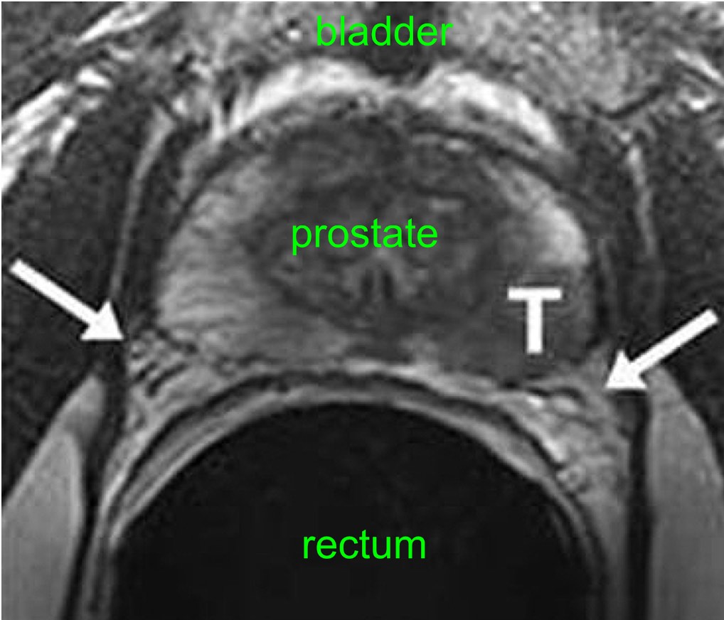
Which tests you might need will depend on the situation. For example, a prostate biopsy is typically done with transrectal ultrasound and/or MRI to help guide the biopsy. If you are found to have prostate cancer, you might need imaging tests of other parts of your body to look for possible cancer spread.
The imaging tests used most often to look for prostate cancer spread include:
Fact Sheet: Molecular Imaging And Prostate Cancer
Prostate cancer is the second leading cause of cancer death in American men, behind only lung cancer. About 1 in 41 men will die of prostate cancer. Prostate cancer is more common in men who are 65 years and older, with an average age at diagnosis of about 66 years.
The American Cancer Society estimates that by the end of 2022, there will be nearly 268,480 new cases of prostate cancer diagnosed in the United States, and about 34,500 will die of the disease.
When detected early, prostate cancer has more than a 95 percent cure rate. Screening for prostate cancer is performed with a simple blood test measuring PSA level and starts at the age of 55. Screening may start as early as the age of 40 for men with increased risk factors. Once prostate cancer is diagnosed, treatment may be highly individualized, and molecular imaging technologies dramatically improve how prostate cancer is localized and treated.
Treatment options include surgery to remove the prostate, radiation therapy, and hormonal or chemotherapy. Determining whether prostate cancer has spread to the lymph nodes or other parts of the body is critical for making appropriate decisions on whether and how to treat prostate cancer. In addition to improving the accuracy of prostate cancer diagnosis, molecular imaging tools can provide detailed information about the cancer that helps patients and their physicians choose the best treatment option.
What is molecular imaging, and how does it help people with prostate cancer?
Recommended Reading: How To Cure Inflamed Prostate
Current And Future Potential Uses For Mpmri
mpMRI of the prostate has the potential to fulfil multiple roles in regard to prostate cancer, including:
-Improving diagnostic accuracy -Post-op follow-up for cancer recurrence -Characterisation of prostatic tissue -and more recently, guidance for targeted biopsy
As Radiologists we recognise that management of patients with suspicion of prostate cancer can be very complex and we want to provide mpMRI on correctly selected patients for specific outcomes. South Coast Radiology will continue to work closely with local Urologists to ensure that the examination is useful in assisting patient management. As such we will only be providing mpMRI prostate from a Urologists referral.
mpMRI can also be helpful to the Urologists who are managing patients under active surveillance protocols. These are patients who are considered low-risk, but need to be monitored for detection of cancer progression.
Fda Approves New Imaging Drug For Detecting Spread Of Prostate Cancer
https://www.facingourrisk.org/XRAY/new-imaging-for-prostate-cancer
Full article: https://www.fda.gov/news-events/press-announcements/fda-approves-first-psma-targeted-pet-imaging-drug-men-prostate-cancer
On December 1, 2020 the FDA approved a new type of imaging technology to confirm the spread of newly diagnosed prostate cancer that is suspected to be metastatic. The approval also includes use for confirming suspected recurrence in men who have rising PSA after treatment. The approval is based on two clinical trials that showed this new technique to be safe and consistent in accurately detecting cancer that has spread beyond the prostate gland.
THIS INFORMATION HAS BEEN UPDATED on 5/10/2022: On March 23, 2022 the U.S. Food and Drug Administration approved a new drug called Pluvicto to treat patients with metastatic castration-resistant prostate cancer. ON the same day, the FDA also approved a new imaging drug called Locametz for identification of those patients who would benefit from treatment with Pluvicto. Read about the FDA approval of Pluvicto and Locametz here.
Read Also: What Is A Transrectal Ultrasound Of The Prostate
How To Receive A Psma Pet Scan And Prostate Cancer Care At Ucla Health
UCLA Health provides comprehensive and customized care for men with prostate cancer. Our physicians use the PSMA PET scan alongside radiation therapy, chemotherapy, surgery, and all other treatment modalities offered to make sure men receive the best possible care from diagnosis to treatment to follow-up.
To ensure the best treatment possible, UCLA Healths nuclear medicine physicians, medical oncologists, radiation oncologists, radiologists, urologists and surgeons optimize care for each person receiving treatment. The prostate cancer care at UCLA Health is backed by multidisciplinary tumor boards with physicians from different specialties and subspecialties, along with genetic counselors and representatives from allied health services. Together, these specialists and experts meet once a week to talk about each new cancer patient and the path forward for their specialized treatment.
When You Need Themand When You Dont
It is normal to want to do everything you can to treat prostate cancer. But its not always a good idea to get all the tests that are available. You may not need them. And the risks from the tests may be greater than the benefits.
The information below explains why cancer experts usually do not recommend certain imaging tests if you are diagnosed with early-stage prostate cancer. You can use this information to talk about your options with your doctor and choose whats best for you.
How is prostate cancer usually found?
Prostate cancer is cancer in the male prostate gland. It usually grows slowly and does not have symptoms until it has spread. Most men are diagnosed in the early stages when their doctor does a rectal exam or a PSA blood test. PSA is a protein made in the prostate. High levels of PSA may indicate cancer in the prostate.
If one of these tests shows that you might have prostate cancer, you will be given more tests. These tests help your doctor find out if you actually have cancer and what stage your cancer is.
What are the stages of prostate cancer?
Prostate cancer is divided into stages one to four . Cancer stages tell how far the cancer has spread.
Stages I and II are considered early-stage prostate cancer. The cancer has not spread outside the prostate. However, stage II cancer may be more likely to spread over time than stage I cancer. In stages III and IV, the cancer has already spread to other parts of the body.
Imaging tests have risks.
09/2012
Also Check: What Is The Female Prostate
Newly Approved Radiotracers Improve Pet/ct Imaging For Prostate Cancer
The FDA approved two radiotracers in that are used with PSMA PET. Gallium 68 PSMA-11 was approved in December, 2020, and the more widely available Pylarify was FDA approved in May, 2021.
Clinical trials showed that PSMA PET found significantly more cancer than conventional imaging methods. In one trial, the new imaging led to a change in the treatment plan for 64% of the participants.
PSMA PET/CT is an imaging tool that uses positron emission tomography fused with computed tomography to detect prostate cancer anywhere in the body. A safe, radioactive substance called a radiotracer is injected into your body before the imaging test. The radiotracer seeks out and attaches to cancer cells, making them visible on the image. In the case of PSMA PET, the radiotracer attaches to a protein on the surface of prostate cancer cells, called prostate specific membrane antigen .
A board-certified physician who specializes in PET/CT evaluates the images and a doctor who specializes in cancer treatment uses the information to develop and monitor cancer treatment.
Disease Recurrence After Treatment

Biochemical recurrence occurs in 20%â40% of patients within 10 y of âdefinitiveâ PCa therapy, often preceding clinically detectable disease . Accurate delineation of local versus metastatic disease is imperative to determine appropriate therapy. Although MRI is widely used to assess local recurrence, its interpretation can be confounded by inherently low specificity and by the glandular atrophy and fibrosis induced by radiation. With respect to PET, PCa grows slowly, accounting for its lack of avidity for 18F-FDG, which has proved so successful for most other malignancies. Also, the bladder produces a strong 18F-FDG signal near the prostate. The role of 11C-choline, as previously described, is limited in detecting early recurrence by its inability to identify microscopic foci of metastatic PCa . Although higher urinary excretion of the fluorinated analogs represents a disadvantage in imaging PCa, 18F-fluorocholine PET/CT has demonstrated a sensitivity of 71% in localizing recurrent disease . 11C-Acetate has also been evaluated for detection of recurrence and has demonstrated results comparable to those of 11C-choline .
Imaging PCa using urea-based low-molecular-weight agents. Dual-pinhole SPECT/CT of PC-3 PIP and PC-3 flu tumor-bearing mouse with +. Note lack of radiopharmaceutical out-side of the tumor. .)
Read Also: Why Does The Prostate Get Enlarged
Improved Technology For Identifying Metastatic And Recurrent Prostate Cancer
Priti Patel, CNMT, left, and Terence Wong, MD, PhD, right, meet with a patient before his PSMA PET/CT scan.
A new imaging technique is changing the way aggressive prostate cancer is identified and is making it easier for doctors to design more effective, individualized treatment plans. Duke Health was one of the first centers in the U.S. to offer prostate specific membrane antigen PET/CT imaging following FDA approval of a new radioactive tracer in May 2021. The new technology can identify cancer both in and outside the prostate gland and especially benefits men whose cancer has recurred and are at risk for it spreading to other parts of the body, even after previous treatments.
Who Interprets The Results And How Do I Receive Them
A Consultant Radiologist, a Doctor specifically trained to interpret radiology examinations, will analyse the images and send a report to your referring Doctor
#prostatecancer #ProstateMRI #prostatecancerawareness #lungcancer #BreastUltrasound #ultrasound #Dexa, #BackMRI #breastcancerawareness #mammogram #AmimagingNJ #advancedwomenimagingnj #breastMri #AdvancedMagneticImaging. #westnewyorknj #unioncitynj #northbergennj #Hoboken #guttenbergnj #MRI
For more information visit. www.advancedimagingnj.com
Follow us in facebook, instagram, tweeter @AMimagingNJ
You May Like: Metastatic Castration Resistant Prostate Cancer Life Expectancy
Lymph Node Biopsy As A Separate Procedure
A lymph node biopsy is rarely done as a separate procedure. Its sometimes used when a radical prostatectomy isnt planned , but when its still important to know if the lymph nodes contain cancer.
Most often, this is done as a needle biopsy. To do this, the doctor uses an image to guide a long, hollow needle through the skin in the lower abdomen and into an enlarged node. The skin is numbed with local anesthesia before the needle is inserted to take a small tissue sample. The sample is then sent to the lab and looked at for cancer cells.
Current Diagnostic Testing For Prostate Cancer
Currently, screening for prostatic adenocarcinoma is based on two tests digital rectal examination and prostate-specific antigen . If either of these are abnormal, a transrectal ultrasound guided biopsy is often the next step and prostate cancer diagnosis is typically made through TRUS-guided sextant biopsy and histopathological examination. However, each of these tests has limitations DRE has low overall sensitivity , and PSA measurement has higher detection rates but the specificity is low , from false-positive PSA elevation under benign circumstances. TRUS biopsy also has limitations as random biopsy sampling can miss the tumour entirely, or the most aggressive part.
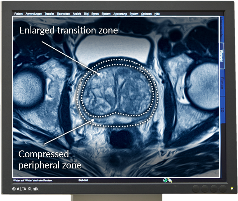
Urologists also often face the dilemma of managing patients who have a high degree of suspicion for cancer and a pathological diagnosis cannot be confirmed. In these patients mpMRI can help as it has the potential to identify suspicious areas within the prostate so that TRUS biopsy can be targeted to this zone.
Don’t Miss: What Does A Nodule On The Prostate Mean
Additional Pet Scanning Faq
Is PET scanning safe?
While PET scanning does involve the use of radioactive tracers, these are diagnostic levels of radiation that are completely safe and have no known side effects.
How long does it take to get the results after a PET Scan?
Images are captured and created during the PET scan procedure, and afterward, a radiologist will utilize their training and experience to interpret the scan and produce a written report of findings and conclusions. This report is then transmitted to your referring physician who would then review those results with you and discuss any further treatment if needed.
What is it like for a patient to have a PET Scan?
Please watch this video we created that clearly walks one through the experience of having a PET scan and answers all common questions about PET scanning.
Positron Emission Tomography Scan
A PET scan is similar to a bone scan, in that a slightly radioactive substance is injected into the blood, which can then be detected with a special camera. But PET scans use different tracers that collect mainly in cancer cells. The most common tracer for standard PET scans is FDG, which is a type of sugar. Unfortunately, this type of PET scan isnt very useful in finding prostate cancer cells in the body.
However, newer tracers, such as fluciclovine F18, sodium fluoride F18, and choline C11, have been found to be better at detecting prostate cancer cells.
Other newer tracers, such as Ga 68 PSMA-11, 18F-DCFPyl , and Ga 68 gozetotide , attach to prostate-specific membrane antigen , a protein that is often found in large amounts on prostate cancer cells. Tests using these types of tracers are sometimes referred to as PSMA PET scans.
These newer types of PET scans are most often used if its not clear if prostate cancer has spread. For example, one of these tests might be done if the results of a bone scan arent clear, or if a man has a rising PSA level after initial treatment but its not clear where the cancer is in the body. PSMA PET scans can also be used to help determine if the cancer can be treated with a radiopharmaceutical that targets PSMA.
Doctors are still learning about the best ways to use these newer types of PET scans, and some of them might not be available yet in all imaging centers.
Read Also: What’s A Prostate Exam
