Prostate Mri Can Help You Avoid Unnecessary Biopsy
Prostate biopsies are used to confirm cancer in high-risk patients suspected to have aggressive prostate cancer. Uncomfortable and invasive, men undergoing the procedure are extremely prone to complications like antibiotic-resistant infections and sepsis. Its estimated that 18% of patients experience some sort of complication, while as many as 4% develop an infection requiring hospital care.
27% of the one million prostate biopsies performed each year are unnecessary.
Typically, a biopsy will be recommended to a patient for one of two reasons: they tested high for levels of the PSA protein, or, the results from a digital rectal exam show they may have prostate cancer. The issue here is that PSA tests are not always accurate.
A males PSA level can be affected by a number of other factors, such as recent sexual activity, an enlarged prostate, and prostatitis. Even a long bicycle ride can cause levels to spike. This leaves a lot of room for false-positives – which lead to unnecessary biopsies.
What Are The Risks Of Breast Mri
A breast MRI is a safe procedure that doesnt expose you to radiation. But as with other tests, a breast MRI has risks, such as: 1 False-positive results. A breast MRI may identify suspicious areas that, after further evaluation such as a breast ultrasound or breast biopsy turn out to be benign. These results are known as false-positives. A false-positive result may cause unneeded anxiety if you have additional testing, such as a biopsy, to assess the suspicious areas. 2 Reaction to the contrast dye used. A breast MRI involves injection of a dye to make the images easier to interpret. This dye can cause allergic reactions and can cause serious complications for people with kidney problems.
Can Mri Help Detect Cancer Yes Heres How
Lets explain the answer behind can MRI detect cancer.
When the technologist pulses a radiofrequency current through your body in the MRI scanner, it stimulates the protons in soft tissues and organs, such as water and fat content at the molecular level.
As a result, the protons move against the magnetic field. When the radiofrequency current ceases, the MRI machines sensors detect the energy the protons expended as they came into alignment with the magnetic field.
While the radio waves are pulsing on and off, the pictures they create allow the radiologist and your oncologist to read the time it took for the protons to realign and how much energy they released.
These differences help determine the molecules chemical signature and identify cancerous tumors and masses.
Also Check: Is Garlic Good For Prostate Enlargement
When To Order An Mri In The Initial Evaluation And Management Of Prostate Cancer
This article summarizes the current role of multiparametric MRI in the diagnosis, risk assessment, and treatment of prostate cancer.
Prostate cancer remains the only solid tumor diagnosed using transrectal ultrasoundguided sampling of the gland, and not an image-based, lesion-directed approach. This technique has limitations in that it underdiagnoses clinically significant disease and overdiagnoses indolent tumors resulting in overtreatment of patients. Technical advances in MRI in the last decade have made this method the preferred imaging modality for prostate anatomy and for risk assessment of prostate cancer. As of 2018, the indications for MRI in the diagnosis and risk assessment of prostate cancer have expanded from preoperative evaluation to the pre-biopsy setting, as well as for surveillance protocols. This article summarizes the current role of multiparametric MRI in the diagnosis, risk assessment, and treatment of prostate cancer.
Mri For Prostate Cancer Screening

In the United States, MRI is primarily used to assist with prostateThe prostate is a walnut-sized gland located between the bla… Full DefinitioncancerCancer is a group of diseases where cells grow abnormally an… Full Definition grading and staging. However, there is great interest in exploring MRI for initial prostate cancer screening as well. MRI for screening has proven popular in Europe, where more studies have been conducted to determine its accuracy in spotting tumors. According to a 2015 study, adding prostate MRI to prostate screening improves cancer detection and reduces unnecessary biopsies. This is an exciting possibility that could help many men avoid the discomfort and side effects of biopsyAn examination of tissue removed from the body to understand… Full Definition.
While there is a lot of evolving data, pre-biopsy MRI is not yet the diagnostic standard in the U.S., which means doctors are not likely to recommend it . This is one of those instances where you should ask your doctor if prostate MRI prior to biopsy might be helpful in your case. There may be clinical trials you can participate in or guidelines may change based on new research. Its always okay to ask for different options.
Read Also: What Does It Mean To Have An Enlarged Prostate
Use In Men Already Diagnosed With Prostate Cancer
The PSA test can also be useful if you have already been diagnosed with prostate cancer.
- In men just diagnosed with prostate cancer, the PSA level can be used together with physical exam results and tumor grade to help decide if other tests are needed.
- The PSA level is used to help determine the stage of your cancer. This can affect your treatment options, since some treatments are not likely to be helpful if the cancer has spread to other parts of the body.
- PSA tests are often an important part of determining how well treatment is working, as well as in watching for a possible recurrence of the cancer after treatment .
How Many Women Have Breast Cancer
One in eight women will be diagnosed with breast cancer, making it the most common cancer for women after skin cancer. Thankfully, breast cancer treatments have come a long way, so more women can enjoy life long after receiving a breast cancer diagnosis. A major factor in the success of breast cancer treatment is early detection.
Also Check: Zytiga For Metastatic Prostate Cancer
What Are The Advantages And Disadvantages Of Having An Mri Scan Before A Biopsy
Advantages
- It can give your doctor information about how likely it is that you have cancer in your prostate.
- Its less likely than a biopsy to pick up a slow-growing cancer. This means you are less likely to have a biopsy and treatments that could have life-changing side effects if your cancer is unlikely to cause you any problems in your lifetime. Some side effects include severe infections and long lasting urinary or sexual problems.
- It can help your doctor decide if you need a biopsy if theres nothing unusual on the scans, this means youre unlikely to have prostate cancer that needs to be treated. You may be able to avoid having a biopsy, and its possible side effects.
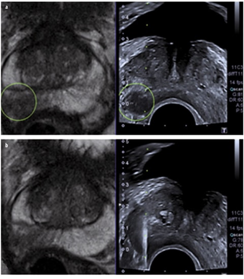
- If you do need a biopsy, your doctor can use the scan images to decide which parts of the prostate to take samples from.
- If your biopsy finds cancer, you probably wont need another scan to check if it has spread, as the doctor can get this information from your first MRI scan. This means you can start talking about suitable treatments as soon as you get your biopsy results.
Disadvantages
- Being in the MRI machine can be unpleasant if you dont like closed or small spaces .
- Some men are given an injection of dye during the scan this can sometimes cause mild side effects.
What Are Some Common Uses Of The Procedure
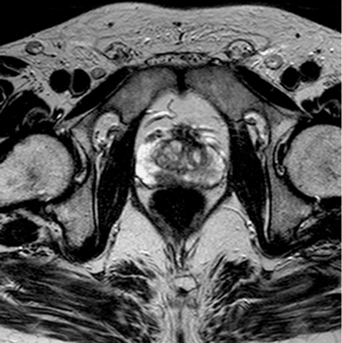
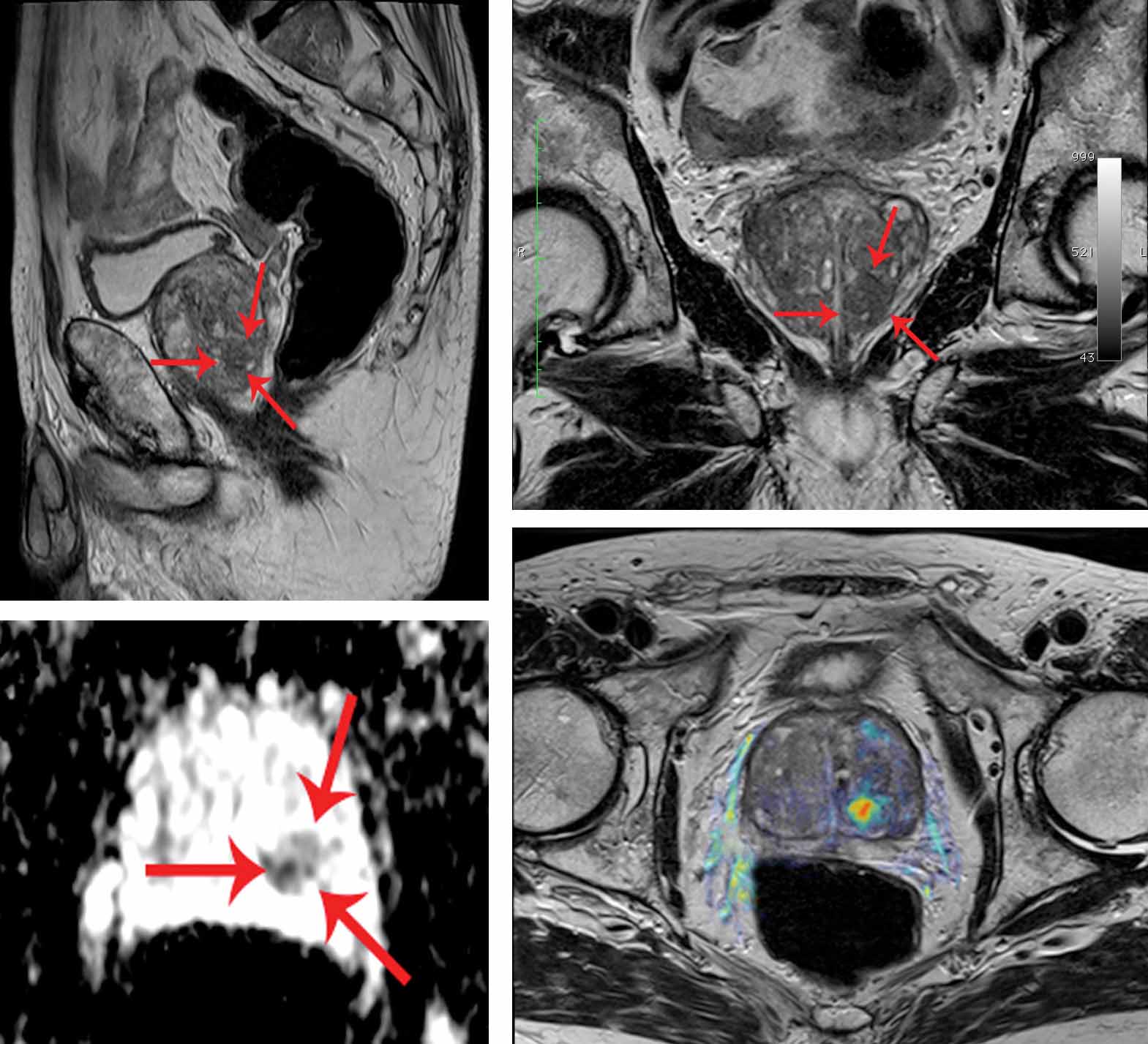
Your doctor uses MRI to evaluate prostate cancer and see if it is limited to the prostate. Mp-MRI provides information on how water molecules and blood flow through the prostate. This helps determine whether cancer is present and, if so, whether it is aggressive and if it has spread.
Occasionally, MRI of the prostate is used to evaluate other prostate problems, including:
- infection or prostate abscess.
Also Check: Can You Climax After Prostate Removal
One Of Our Many Initiatives To Improve Diagnosis
It was thanks to our funding of a pilot study into mpMRI by Hash Ahmed and Mark Emberton at University College London Hospital in 2010, that the research team went on to secure £2 million of Government funding to carry out the PROMIS trial. But mpMRI is just one of the ways were working to improve diagnosis of prostate cancer.
We also funded the PROMIS trials researchers to collect and store blood and urine samples from the men taking part, to see if tests using biomarkers can be developed to improve diagnostic accuracy even further. And weve just launched our Stronger Knowing More campaign to raise awareness among black men of their great risk of prostate cancer.
Its all part of our ongoing strategy to tame the most common cancer in men within ten years.
What Is Colorectal Cancer
Colorectal cancer is a disease in which abnormal cells in the colon or rectum divide uncontrollably, ultimately forming a malignanttumor.
Most colorectal cancers begin as a growth, or lesion, in the tissue that lines the inner surface of the colon or rectum. Lesions may appear as raised polyps, or, less commonly, they may appear flat or slightly indented. Raised polyps may be attached to the inner surface of the colon or rectum with a stalk , or they may grow along the surface without a stalk .
Colorectal polyps are common in people older than 50 years of age, and most do not become cancer. However, a certain type of polyp known as an adenoma is more likely to become a cancer.
Colorectal cancer is the third most common type of non-skin cancer in both men and women . It is the second leading cause of cancer death in the United States after lung cancer. In 2021, an estimated 149,500 people in the United States will be diagnosed with colorectal cancer and 52,980 people will die from it .
Also Check: What Age Do You Start Prostate Screening
Why You Might Have A Mpmri
It is important to know that a mpMRI scan alone cannot diagnose prostate cancer. But it can help your doctor to see:
- whether you have an area in the prostate gland that looks suspicious
- if you need a biopsy
- where to take the biopsies from your prostate
- if the suspected cancer is just within the prostate or has spread outside it
What Happens If You Have A Likert Score Of 1 Or 2

Your doctor may not recommend a biopsy if you have a low Likert score. But you can still have one if you want to. Your doctor will explain the possible benefits and risks of having a biopsy.
When recommending whether you need a biopsy or not, your doctor also looks at other factors. These include:
- other test results such as PSA level and prostate examination
- the size of your prostate and the corrected PSA for its size. This is called the PSA density
- any other health conditions that you might have
- how well youd cope with prostate cancer treatment and whether it would benefit you
They might decide not to recommend a biopsy if you’re unwell or not likely to be able to have treatment.
If you don’t have a biopsy, your doctor may recommend monitoring your PSA level. They will recommend what your PSA level should be. You are usually discharged back to your GP to be monitored. Your GP can refer you back if the PSA levels go up.
You May Like: How To Tell Prostate Cancer
What Are The Risks Of Breast Cancer
Here are some of the factors that can increase a womans risk of breast cancer: 1 More than two close relatives with breast cancer 2 A close family member diagnosed with breast cancer at an early age 3 Having a mutation in a gene linked to breast cancer , or having a family member with the mutation 4 A personal history of an abnormal breast biopsy
What Is Breast Mri
A breast MRI is a specific type of MRI that provides detailed images of the breast. Breast MRI machines have special breast coils that help create high-resolution and high-quality images of one or both breasts. These MRIs can create detailed, three-dimensional images of breast tissue, allowing radiologists to better identify any abnormalities.
Don’t Miss: How Long Will Hormone Therapy Work For Prostate Cancer
Is Mri Examination Safe
Yes. The MRI examination poses no risk to the average patient if appropriate safety guidelines are followed.
People who have had heart surgery and people with the following medical devices can be safely examined with MRI:
- Surgical clips or sutures
- Weight of more than 300 pounds
- Not being able to lie on back for 30 to 60 minutes
Statistical And Cost Analysis
We report the proportions of patients in each risk group who received a bone scan and subsequently underwent imaging and procedures. We further explore regional variation in use, as well as variation by primary treatment modality . The reason for the latter is that external beam radiation often includes a CT and/or MRI scan for treatment planning, which can be a source of confounding in this analysis for these 2 specific categories of imaging. Multivariate logistic regression models assessed for covariates associated with bone scan use in each risk group. All analysis was performed with SAS 9.2 , and a P value of < .05 was considered statistically significant.
Direct cost to Medicare was calculated based on the cost of a bone scan from the CPT code 78306, which accounted for the majority of Medicares bone scan claims among patients with prostate cancer. The 2012 Medicare national average reimbursement rate of $257.66 was multiplied by the estimated annual incidence of prostate cancer in patients older than 65. This figure was then multiplied by the proportion of patients receiving bone scans as reported in this study. Similarly, the costs of downstream imaging and procedures were estimated using 2012 national reimbursement rates multiplied by the relative frequency of each procedure.
Don’t Miss: Can Sitting For Long Periods Cause Prostatitis
How Does Mri Work
An MRI scanner is a long cylinder or tube that holds a large, very strong magnet. You lie on a table that slides into the tube, and the machine surrounds you with a powerful magnetic field. The machine uses a powerful magnetic force and a burst of radiofrequency waves to pick up signals from the nuclei of hydrogen atoms in your body. A computer converts these signals them into a black and white picture.
Contrast materials can be put into the body through a vein to make the images clearer. Once absorbed by the body, the contrast speeds up the rate at which tissue responds to the magnetic and radio waves. The stronger signals give clearer pictures.
Biopsy During Surgery To Treat Prostate Cancer
If there is more than a very small chance that the cancer might have spread , the surgeon may remove lymph nodes in the pelvis during the same operation as the removal of the prostate, which is known as a radical prostatectomy .
The lymph nodes and the prostate are then sent to the lab to be looked at. The lab results are usually available several days after surgery.
Read Also: Should I Have My Prostate Removed
How It Works And What To Expect
Most scans donât take more than an hour or so, though you may have to wait a few hours as health care workers prep you for the test.
These scans are usually done at a nuclear medicine or radiology department at a hospital. A little bit of radioactive material will go into your body. Doctors call this material a radioactive âtracer,â radionuclide, or radiopharmaceutical.
Hospital staff may inject this tracer or give it to you to swallow in a pill or inhale as a gas.
It can take from a few seconds to several days for the tracer to collect in the part of the body that will get scanned.
Before the scan, youâll remove all jewelry and metal that could interfere with the images. Medical staff may ask you to wear a hospital gown, though in some cases you can wear your own clothes.
Youâll lie on a table or sit on a chair for the scan. Technicians use a special camera, or âscanner,â on the appropriate parts of your body to detect gamma rays from the tracer. Technicians might ask you to change positions to get different angles as the scanner works.
The scanner sends the information to computer software that creates pictures, sometimes in three dimensions and with color added for clarity.
A specialized doctor called a radiologist will review the pictures and talk to your doctor about what they show.
Recommended Reading: Docetaxel Prostate Cancer Life Expectancy
What Are The Possible Complications Of An Mri

People can be hurt in MRI machines if they take metal items into the room or if other people leave metal items in the room.
Some people become very uneasy and even panic when lying inside the MRI scanner.
Some people react to the contrast material. Such reactions can include:
- Pain at the needle site
- A headache that develops a few hours after the test is over
- Low blood pressure leading to a feeling of lightheadedness or faintness
Be sure to let your health care team know if you have any of these symptoms or notice any other changes after you get the contrast material.
Gadolinium, the contrast material used for MRI, can cause a special complication when its given to patients on dialysis or who have severe kidney problems, so its rarely given to these people. Your doctor will discuss this with you if you have severe kidney problems and need an MRI with contrast.
Small amounts of gadolinium can stay in the brain, bones, skin and other parts of your body for a long time after the test. Its not known if this might have any health effects, but so far, studies havent found any harmful effects in patients with normal kidneys.
Recommended Reading: What Is The Best Prostate Supplement On The Market
