Prostate Biopsy Specimens: Ask For Meticulous Labeling
The way that prostate biopsy samples are handled varies among hospitals. The samples, or “cores,” are put onto glass slides to be examined under a microscope for signs of cancer. Find out if the physician doing the biopsy will place each core in a separate, labeled container. If cancer is discovered, its location in the prostate gland can affect decisions about further testing and possible treatment.
Individual labeling of biopsy cores is more expensive, and not all hospitals provide this level of service. “If all of the samples from the right and left side of the prostate gland are processed together, as opposed to individually, consider having the biopsy done elsewhere,” Dr. Garnick says.
What If My Mri Prostate Biopsy Is Positive
Should your MRI fusion biopsy indicate the presence of prostate cancer, Dr. Kasraeian will discuss all of your options, including further evaluation, active surveillance, and prostate cancer treatment. The most appropriate course of action will depend on the nature, severity, and location of the prostate cancer, as well as the patients age, personal wishes, whether the cancer has metastasized, and other factors. Kasraeian Urology offers a number of advanced and effective prostate cancer treatment options, including:
- Robotic prostatectomy
What Will I Experience During And After The Biopsy
If you receive IV contrast for the MRI-guided procedure, you may feel coolness and a flushing sensation for a minute or two following the injection. The IV needle may cause some discomfort when the doctor inserts it and you may have some bruising when they remove it.
Rarely, patients may experience side effects from MR contrast material, such as nausea and local pain, hives, itchy eyes, or other reactions. If you have allergic symptoms, a radiologist or other doctor will be available for immediate assistance.
When the doctor inserts the ultrasound probe or endorectal coil into the rectum, you will feel pressure and may have some temporary discomfort.
You will hear a clicking noise when the biopsy needle samples the prostate and you may feel a stinging or burning sensation in the area.
Some patients find it uncomfortable to remain still during an MRI. Others experience a sense of being closed-in . Sedation is available for patients who anticipate anxiety.
If you feel heating on your skin at any time during MR imaging, tell the MR technician so they can closely examine the area.
Some patients experience a small amount of bleeding from the rectum or perineum immediately after the biopsy. If this does occur, it will cease with gentle pressure.
If you did not receive sedation, no recovery period is necessary. Light general anesthetic or sedation may leave you feeling groggy for a day or so.
Read Also: Support Group For Prostate Cancer
Koelis Technology For Fusion Biopsy
For over a decade KOELIS® has assisted urologists and radiologists from around the world in their routine clinical practice providing the latest technology for personalized prostate cancer.
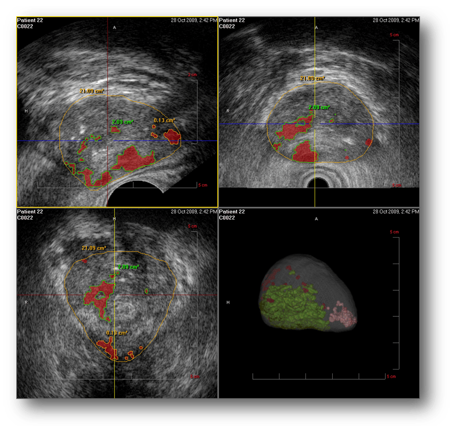
We propose precise real-time 3D transrectal ultrasound fusion-guided transperinal and transrectal prostate biopsy against conventional and random systematic biopsy to increase sampling quality with KOELIS Trinity®. In a simple process and device, KOELIS Trinity® cartographer is a powerful diagnostic tool to plan, implement, review and control a personalized care solution. It creates a detailed and personalized map of the patients prostate showing accurate core distribution since our image-based cartographer is equipped with the Organ-Based Tracking® technology. Without changing the usual clinical practices, this technique brings an increased quality control over biopsy localizations. A precise, individual prostate biopsy mapping is a value for the accurate diagnosis and the further management of each patient.
Health innovation as a passion
At KOELIS®, we innovate every day in collaboration with world-renowned universities and hospitals to offer physicians new advancements in imaging and a greater field of view in order to bring personalized answers to every patient, in the respect of their quality of life.
Do you have questions about our precision fusion technology? Find the common inquiries or contact us !
Read Also: How To Reach Your Prostate
Leading Specialists In Laser Ablation Procedure For Prostate Tumors

Are you, or is someone you love, at risk for prostate cancer? Have you been told that your PSA test results are high and you need a biopsy? Have you had a positive biopsy? Are you exploring alternatives to surgery or radiation?
The world of prostate cancer can be overwhelming, with confusing and conflicting information about the disease and its treatments, but little explaining what is happening inside your own body.
Rest assured. The Sperling Prostate Center team is here to put your mind at ease.
Patient Greyson Quarles tells his story
The Sperling Prostate Center in Florida offers noninvasive advanced imaging as a revolutionary first step in prostate tumor detection and diagnosis. The more you know about what is happening in your body, the more informed and rational your healthcare decisions will be. The best education is having a clear picture of what is going on within your own body. Dr. Sperling and his team will welcome and support you as advanced images are captured, your prostate gland is mapped, and the results are explained to you in a way you can fully understand.
Recommended Reading: Blood Test For Prostate Cancer Levels
What Happens After A Prostate Biopsy
Your recovery process will vary depending on the type of anesthesia that isused. If you were given general anesthesia, you will be taken to a recoveryroom for observation. Once your blood pressure, pulse, and breathing arestable and you are alert, you will be taken to your hospital room ordischarged to your home.
If local anesthetic was used, you may go back to your normal activities anddiet unless otherwise instructed. You may feel the urge to urinate or havea bowel movement after the biopsy. This feeling should pass after a fewhours.
There may be blood in your urine or stool for a few days after the biopsy.This is common. Blood, either red or reddish brown, may also be in yourejaculate for a few weeks after the biopsy. This, too, is normal.
The biopsy site may be tender or sore for several days after the biopsy.Take a pain reliever for soreness as recommended by your healthcareprovider. Aspirin or certain other pain medicines may increase the chanceof bleeding, so be sure to take only recommended medicines.
-
Increase in the amount of blood in your urine or stool
-
Belly or pelvic pain
-
Trouble urinating
-
Changes in the way your urine looks or smells or burning with urination
-
Fever and/or chills
Your healthcare provider may give you other instruction, depending on yoursituation.
Offers Extra Benefits For Prostate Cancer Follow
This type of biopsy is advantageous in patients who require a repeat biopsy. They are more likely to tolerate the repeated procedure.
These patients sometimes need to be reclassified when cancer progresses.
A new classification means that the tumor is in a new stage, and the healthcare provider will adjust the treatment.
MRI-guided biopsies detect these changes more accurately, leading to a higher reclassification rate.
Read Also: Can A Man Have Sex If His Prostate Is Removed
What Are The Symptoms Of Prostate Dysfunction
Not all prostate cancer patients experience prostate symptoms. However, according to the National Institute on Aging, the most common symptoms of prostate problems include:
- Increased frequency of urination
- Sexual dysfunction
- Painful back, hips, pelvis, or rectum
If you are experiencing these symptoms, please seek medical care as soon as possible. They may indicate inflammatory conditions like BPH , prostatitis , an infection of the prostate, bladder, or other organs, or other diseases like STDs and cancer.
Lymph Node Biopsy As A Separate Procedure
A lymph node biopsy is rarely done as a separate procedure. Its sometimes used when a radical prostatectomy isnt planned , but when its still important to know if the lymph nodes contain cancer.
Most often, this is done as a needle biopsy. To do this, the doctor uses an image to guide a long, hollow needle through the skin in the lower abdomen and into an enlarged node. The skin is numbed with local anesthesia before the needle is inserted to take a small tissue sample. The sample is then sent to the lab and looked at for cancer cells.
Also Check: Can Losing Weight Shrink Prostate
How To Make Your Prostate Biopsy Go Better
|
Before a prostate biopsy, discuss all thesteps you or your doctor can take to makethe experience as comfortable, safe, andinformative as possible. Image: Wavebreakmedia Ltd/Getty Images |
Here is what men need to know to minimize discomfort of a prostate biopsy and get the best results.
Many men choose to have prostate-specific antigen blood tests to check for hidden prostate cancer, despite the uncertain benefits. Having an abnormal result often leads to a prostate biopsythe only way to confirm the presence of cancer. Biopsies are invasive, but they have become routine.
To reduce discomfort and get the best results, discuss the procedure in detail with your doctor. Certain practices can improve the overall outcomefor example, make sure you get a shot of anesthetic into the prostate to numb pain during the procedure. “Local anesthesia makes a world of difference between having a tolerable biopsy experience and an unpleasant one,” says Dr. Marc B. Garnick, Gorman Brothers Professor of Medicine and a prostate cancer expert at Harvard-affiliated Beth Israel Deaconess Medical Center.
How Do You Identify The Core Before Biopsy
What makes targeting possible is multiparametric MRI .
Prior to biopsy, an mpMRI scan excels at identifying significant prostate cancer. An experienced radiologic reader assigns it a PI-RADS score from 1 to 5, where category 5 indicates the greatest probability of cancer. Thus, a category of 4 or 5 strongly suggests the presence of significant prostate cancer. These are the key cells to target and capture.
When analyzed in a lab, they are given a Grade Group from 1 to 5, where GG 2 indicates significant prostate cancer is present.
Don’t Miss: Can Prostate Cancer Cause Back Pain
Pioneers In Imaging Technology Utilization
Loyola University Medical Center is the first hospital in Illinois to use UroNav®, an MR/TRUS imaging system for patients with suspected prostate cancer.
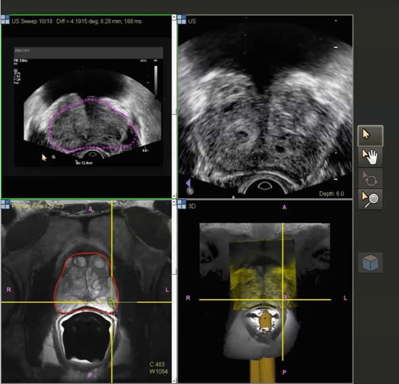
Expert urologic oncologists at Loyola use UroNav technology to fuse pre-biopsy images from magnetic resonance imaging with ultrasound-guided biopsy images in real time to create a detailed, three-dimensional view of the prostate.
An improvement to a standard biopsy, this imaging system can better target areas of the prostate that are hard to see, leading to fewer biopsies, more accurate diagnosis and better treatment decisions for prostate cancer patients.
What Does An Elevated Psa Mean

The PSA test, which detects the level of prostate-specific antigen in a mans blood, is a valuable screening tool for prostate cancer. While the normal PSA range can vary based on a patients age and other factors, a PSA of 4.0 ng/mL or higher is generally considered abnormal for men over 60. Most cases of an abnormal or elevated PSA result can be attributed to a benign condition, though a PSA elevation can also be an indicator of prostate cancer. The most common causes of an elevated PSA include:
- Inflamed prostate
- Injury or trauma to the prostate
- Prostate cancer
Depending on the degree of elevation, the patients PSA history, the patients risk for prostate cancer, and other factors, further diagnostic testing and/or biopsy may be required to rule out disease. Kasraeian Urology is proud to offer the most advanced prostate cancer screening tools available in Greater Jacksonville, including:
- MRI/US fusion biopsy
Recommended Reading: Can A Prostate Biopsy Cause Problems
Open Access License / Drug Dosage / Disclaimer
This article is licensed under the Creative Commons Attribution-NonCommercial-NoDerivatives 4.0 International License . Usage and distribution for commercial purposes as well as any distribution of modified material requires written permission. Drug Dosage: The authors and the publisher have exerted every effort to ensure that drug selection and dosage set forth in this text are in accord with current recommendations and practice at the time of publication. However, in view of ongoing research, changes in government regulations, and the constant flow of information relating to drug therapy and drug reactions, the reader is urged to check the package insert for each drug for any changes in indications and dosage and for added warnings and precautions. This is particularly important when the recommended agent is a new and/or infrequently employed drug. Disclaimer: The statements, opinions and data contained in this publication are solely those of the individual authors and contributors and not of the publishers and the editor. The appearance of advertisements or/and product references in the publication is not a warranty, endorsement, or approval of the products or services advertised or of their effectiveness, quality or safety. The publisher and the editor disclaim responsibility for any injury to persons or property resulting from any ideas, methods, instructions or products referred to in the content or advertisements.
Use In Men Who Might Have Prostate Cancer
The PSA blood test is used mainly to screen for prostate cancer in men without symptoms. Its also one of the first tests done in men who have symptoms that might be caused by prostate cancer.
PSA in the blood is measured in units called nanograms per milliliter . The chance of having prostate cancer goes up as the PSA level goes up, but there is no set cutoff point that can tell for sure if a man does or doesnt have prostate cancer. Many doctors use a PSA cutoff point of 4 ng/mL or higher when deciding if a man might need further testing, while others might recommend it starting at a lower level, such as 2.5 or 3.
- Most men without prostate cancer have PSA levels under 4 ng/mL of blood. Still, a level below 4 is not a guarantee that a man doesnt have cancer.
- Men with a PSA level between 4 and 10 have about a 1 in 4 chance of having prostate cancer.
- If the PSA is more than 10, the chance of having prostate cancer is over 50%.
If your PSA level is high, you might need further tests to look for prostate cancer.
To learn more about how the PSA test is used to look for cancer, including factors that can affect PSA levels, special types of PSA tests, and what the next steps might be if you have an abnormal PSA level, see Screening Tests for Prostate Cancer.
Read Also: What Age To Check For Prostate
Schedule Your Visit With A Uh Urologist Today
To learn more about University Hospitals advanced prostate diagnostic tests, or to schedule an appointment, call us at .
UH is dedicated to advancing men’s health through faster, easier and more accurate prostate cancer diagnosis for reduced anxiety and a better experience overall.
Part of a research grant through the National Institutes of Health , our team at University Hospitals was an important part of this unique discovery which is now available at UH for our patients across Northeast Ohio. Its just one of the ways UH is researching, developing and delivering new and advanced patient care with these MRIs for prostate cancer screening.
What Happens During An Mri
These are done in the outpatient procedure area. Antibiotics will be given to reduce the risk of infection from the biopsy. Your doctor will be using the MRI and ultrasound images to watch where the biopsy needles are going. You may feel some discomfort or mild pain when the ultrasound probe is inserted into the rectum. Local anesthesia is used to ease the discomfort.
Also Check: Side Effects Of Prostate Removal Mayo Clinic
% Less Detection Of Insignificant Cancer 3 Times Less Biopsy Core: Less Complication And Side Effects Mri/us Fusion Cancer Detection Rate
PRECISION study : MRI-Targeted or standard Biopsy for Prostate-Cancer Diagnosis
V.Kasivisvanathanet al. May 2018
- In cases of negative results, the hypothesis of cancer cannot be ruled out.
- In cases of low-positive results, suspicion of more aggressive cancer may remain.
Conventional Biopsy : Possible results
25% of cases Biopsy misses the tumor
Fortuitous reaching of non significant cancer
Underestimation of tumors size
Delaying the diagnosis, under- or overestimating the aggressiveness of the disease and, ultimately, failing to provide the best possible care for the patient.
However, the evolution of medical imaging techniques together with advances in fusion technologies have changed the landscape where practices are concerned.
In technical terms, targeted biopsies are made possible by the fusion of ultrasound images of the prostate with MRI scans or PET sequences, in real time during the procedures.
Recommended Reading: How Is Prostate Test Done
What Does The Equipment Look Like
Ultrasound equipment:
Ultrasound scanners consist of an electronic console containing a computer, video monitor, and a handheld transducer . The transducer sends out inaudible high frequency sound waves into the body and listens for the returning echoes. The principle is similar to the sonar used by boats and submarines.
The computer displays the ultrasound image on a video monitor. This image is based on the amplitude and frequency of the signal. It is also based on signal travel time, tissue composition, and the type of body structure through which the sound travels.
The ultrasound probe for a prostate biopsy is about the size of a finger. Once the doctor inserts the probe into the rectum, they take tissue samples using a spring-driven needle core biopsy device . The handheld device includes a long but very thin needle. The needle opens inside the prostate, takes the sample, and then closes.
MRI equipment:
The traditional MRI unit is a large cylinder-shaped tube surrounded by a circular magnet. You will lie on a table that slides into a tunnel towards the center of the magnet.
Don’t Miss: What Color Represents Prostate Cancer
Welcome To Sperling Prostate Center
Our advanced diagnosis and treatment systems in Florida offer effectiveness and quality of life unattainable with conventional methods. Our patients experience minimal side effects, low recurrence rates, and virtually no risk of incontinence or impotence.
Our MRI-guided detection methods utilize the worlds most advanced 3T MRI system along with multiparametric software to create a vivid, 3D prostate map revealing a 360° view of the gland and any irregularities. This technology allows us to perform more accurate biopsies with up to 80% fewer needle samples than conventional biopsies.
Patients also have access to epigenetic screening that detects DNA changes associated with prostate cancer, making the biopsy more effective and conclusive.
If a tumor is detected, our prostate laser center offers a revolutionary focal laser therapy for prostate cancer that dramatically reduces the risk of impotence and incontinence associated with conventional treatments. This minimally-invasive outpatient procedure eradicates tumors in a single one hour treatment, leaving patients ready for everyday life by the next day. Focal laser therapy also offers a superior therapy for benign prostatic hyperplasia or BPH.
