Screening For Prostate Cancer
Prostate cancer is typically treatable if caught early. More than 90 percent of prostate cancers are found when the disease is in the beginning stages, confined to the prostate and nearby organs.
Unlike screenings for breast and colon cancers, there are no universal screening guidelines for prostate cancer. The U.S. Preventive Services Task Force recommends that men age 55 to 69 weigh the benefits and risks before deciding whether they should undergo screening, which is typically performed with a blood test that measures levels of a protein called prostate-specific antigen .
However, men in high-risk groupssuch as those who are of African-American descent and/or have a first-degree relative diagnosed with prostate cancer before age 65should consider speaking with their doctor about starting screenings at an earlier age.
Men older than 70 shouldnt be routinely screened for prostate cancer, according to the USPSTF.
Regardless of age or risk factors, men should get checked if they suddenly experience issues with urination, erectile dysfunction or unexplained pain.
The USPSTF suggests that, before deciding on a screening, men should seek expert advice about the benefits and harms of screening. Risks may include:
- False positives
- Complications and side effects from biopsies to confirm a diagnosis
- The possibility that a prostate cancer diagnosis wont extend lifespan or improve quality of life
Health History And Physical Exam
Your health history is a record of your symptoms, risk factors and all the medical events and problems you have had in the past.
Your doctor will ask questions about your history of symptoms that suggest prostate cancer, such as changes in bladder habits.
Your doctor may also ask about a family history of:
- prostate cancer
- risk factors for prostate cancer
- other cancers
Your doctor will also do a physical exam to look for any signs of prostate cancer. During a physical exam, your doctor may:
- do a digital rectal exam to check the size and shape of the prostate and feel for any lumps or abnormal areas
- check other areas of your body, including the abdomen
Find out more about physical exams and the digital rectal exam .
Advanced Genomic Testing For Prostate Cancer
The most common lab test for prostate cancer is advanced genomic testing, which examines a tumor to look for DNA alterations that may be driving the growth of the cancer. By identifying the mutations that occur in a cancer cells genome, doctors may get a clearer picture of the tumors behavior and be able to tailor a patients treatment based on the findings.
Get Peace Of Mind With Your Cancer Care
Start Your Journey Today!
Take the first step toward feeling better and having more time with your loved ones: Make sure you have access to the most powerful diagnostic tools and treatments that medical science has to offer with expert guidance from our Scientific Research and Advocacy Team every step of the way.
Imaging Tests For Prostate Cancer

Imaging tests use x-rays, magnetic fields, sound waves, or radioactive substances to create pictures of the inside of your body. One or more imaging tests might be used:
- To look for cancer in the prostate
- To help the doctor see the prostate during certain procedures
- To look for spread of prostate cancer to other parts of the body
Which tests you might need will depend on the situation. For example, a prostate biopsy is typically done with transrectal ultrasound and/or MRI to help guide the biopsy. If you are found to have prostate cancer, you might need imaging tests of other parts of your body to look for possible cancer spread.
The imaging tests used most often to look for prostate cancer spread include:
Abdominal Ct Scan And The Detection Of Bladder Cancer
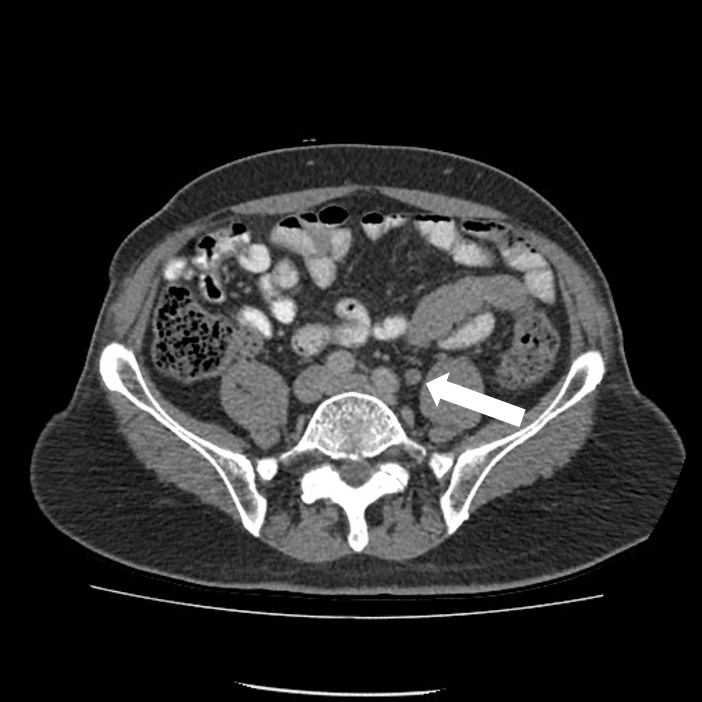
CT scan is able to detect large bladder irregularities, but not always small lesions, says Dana Rice, MD, a board certified urologist and creator of the UTI Tracker mobile app, which helps patients catalog daily urinary tract symptoms, medication and behavioral patterns, and offers personalized tips for UTI prevention.
Also, because the bladder is not a solid organ it is very easy to mistake a bladder fold for an abnormal finding and vice versa, continues Dr. Rice.
For instance, a CT scan may list or read a filling defect which means contrast dye does not fill the bladder evenly.
In this situation the defect may be from many causes, i.e., blood clot, prostate tissues, ureterocele , bladder tumor and more.
Other times, the CT scan can appear completely normal and there may be small tumors not seen or carcinoma in situ . For these reasons, cystoscopy to examine the bladder is the gold standard diagnostic test.
What Concerns Do People Have About Either Imaging Method
For CT, I hear anxiety about exposure to radiation, especially if it is being done repeatedly. For example, certain early-stage cancers can be cured. But you might be coming back every few months or every year for a CT scan. The question is: Do we have an alternative? For detecting cancer that has come back throughout the body, a CT scan is preferable to an MRI. As radiologists, we follow a measure called as low as reasonably achievable. This means we give enough radiation to create CT images that are of high enough quality that we can make a good clinical decision, but we keep the radiation as low as possible to minimize risk.
For MRI, people who have trouble with claustrophobia or are unable to hold their breath, which may be required for certain abdominal imaging tests, may not be able to tolerate the procedure. Some MRI machines can be configured in ways that may reduce claustrophobia. Medical implants, such as a pacemaker, brain stimulator, or other devices, are another complicating factor. The radio waves used with MRI can heat up devices made of metal. This is potentially a concern for something inside the body. Newer medical devices are usually designed with this in mind, so they are safe inside an MRI.
When You Need Themand When You Dont
It is normal to want to do everything you can to treat prostate cancer. But its not always a good idea to get all the tests that are available. You may not need them. And the risks from the tests may be greater than the benefits.
The information below explains why cancer experts usually do not recommend certain imaging tests if you are diagnosed with early-stage prostate cancer. You can use this information to talk about your options with your doctor and choose whats best for you.
How is prostate cancer usually found?
Prostate cancer is cancer in the male prostate gland. It usually grows slowly and does not have symptoms until it has spread. Most men are diagnosed in the early stages when their doctor does a rectal exam or a PSA blood test. PSA is a protein made in the prostate. High levels of PSA may indicate cancer in the prostate.
If one of these tests shows that you might have prostate cancer, you will be given more tests. These tests help your doctor find out if you actually have cancer and what stage your cancer is.
What are the stages of prostate cancer?
Prostate cancer is divided into stages one to four . Cancer stages tell how far the cancer has spread.
Stages I and II are considered early-stage prostate cancer. The cancer has not spread outside the prostate. However, stage II cancer may be more likely to spread over time than stage I cancer. In stages III and IV, the cancer has already spread to other parts of the body.
Imaging tests have risks.
09/2012
What Are Some Disadvantages Of Each Imaging Method
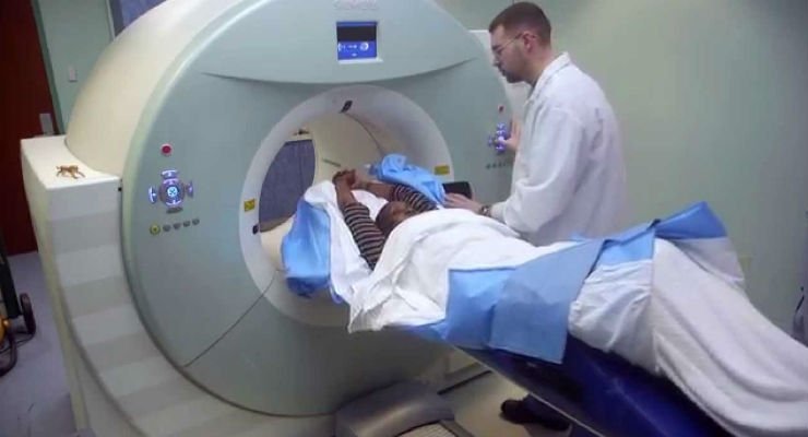
Because CTs use ionizing radiation, they could damage DNA and may very slightly raise the risk of developing cancer. The Food & Drug Administration estimates that the extra risk of any one person developing a fatal cancer from a typical CT procedure is about 1 in 2,000. MRIs do not use ionizing radiation, so there is no issue of raising cancer risk. But they take much longer to complete than CTs. MRIs require the person to lie still within a closed space for about 20 to 40 minutes. This can affect some people with claustrophobia, and the procedure is noisy, which is why we provide ear protection.
Both CT and MRI commonly require the injection of a contrast dye before or during the procedure. This helps the radiologist see organs and other tissues within the body more clearly.
Pet Scanning For Prostate Cancer
If you or a loved one has been diagnosed with any type of cancer, there are often many questions left unanswered. For those diagnosed with prostate cancer, a doctor may suggest having a PET scan as a part of an overall plan for treatment and monitoring for any recurrence or progression of disease. We hope this article may be a resource and help answer a few of the most common questions that we receive regarding PET scanning for prostate cancer.
With prostate cancer being the second leading cause of cancer death in American men , new advances in early detection and high-quality imaging for staging and restaging of prostate cancer have become a top priority for the medical community. Comprehensive cancer care requires high-quality imaging and is the driving force behind rapid and ongoing advancements in imaging techniques used for prostate cancer. In recent years, PET scanning for prostate cancer has become an increasingly popular choice to clearly locate and assess the extent of prostate cancer. While there have been many improvements across many conventional imaging modalities such as TRUS, CT, and MRI, this article is intended to highlight the essential role of advanced imaging such as PET scanning for prostate cancer.
Are you looking to learn more about PSMA PET scanning for prostate cancer? If you are in need of a PSMA PET Scan please join our priority list by
How Can You Protect Yourself
You don’t need to stop getting CT scans. But itâs a good idea to make sure you need each one you get.
Before you have any imaging test, ask your doctor these questions:
- Why do I need this scan?
- How will it affect my treatment?
- What are the risks?
- Could you diagnose me with a test that doesnât use radiation, like an or an ultrasound?
- How will you protect the rest of my body during the CT scan?
Your doctor should use the smallest possible dose of radiation to do the scan — especially if you need to have several of them. Ask if the technician can shield the parts of your body that donât need the test with a lead apron. This will block the X-rays from entering those areas.
Keep a record of the CT scans and other X-rays you have, so you’ll know how much radiation you’re getting. It will also prevent you from repeating a test you’ve already done.
Write down:
Aiding Cancer Detection During Ct Scans
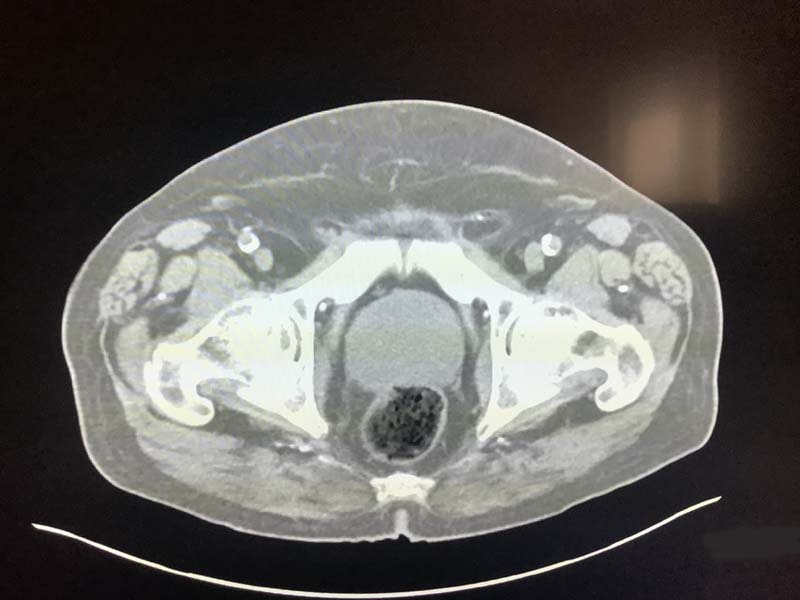
Due to the high doses of radiation involved, CT imaging is not suitable for regular cancer screening. However, the new AI solution could allow for cancer detection whenever men have their abdomen or pelvis examined for other, unrelated issues.
Dr Ruwan Tennakoon, from RMIT University, said that, whilst the CT scans were effective at detecting bone and joint issues, even trained radiologists were unable to spot prostate cancers on the images.
What Else Should I Know About This Test

- Although a CT scan is sometimes described as a slice or a cross-section, no cutting is involved.
- The amount of radiation you get during a CT scan is a good deal more than that with a standard x-ray.
- People who are very overweight may have trouble fitting into the CT scanner.
- Be sure to tell your doctor if you have any allergies or are sensitive to iodine, seafood, or contrast dyes.
- Tell your doctor if you could be pregnant or are breastfeeding.
- CT scans can cost up to 10 times as much as a standard x-ray. You may want to be sure your health insurance will cover this test before you have it.
Our team is made up of doctors and oncology certified nurses with deep knowledge of cancer care as well as journalists, editors, and translators with extensive experience in medical writing.
American College of Radiology/Radiological Society of North America. Body CT/CAT scan. September 23, 2014. Accessed at www.radiologyinfo.org/en/info.cfm?pg=bodyct on November 13, 2015.
American College of Radiology/Radiological Society of North America. Computed Tomography Abdomen and Pelvis. August 13, 2014. Accessed at www.radiologyinfo.org/en/info.cfm?pg=abdominct on November 13, 2015.
American College of Radiology/Radiological Society of North America. Computed Tomography landing page. Accessed at www.radiologyinfo.org/en/submenu.cfm?pg=ctScan on November 13, 2015.
Last Revised: November 30, 2015
When And Why Are Pet Scans Used For Prostate Cancer
- Staging: A PET Scan is used for patients with known prostate cancer in order to pinpoint its exact location and to determine the extent of disease and to determine a treatment strategy.
- Planning Treatment: In some instances, a PET scan may be used to specifically target certain high-risk areas for special treatment. There are occasions when a PET scan is the only diagnostic test that can identify these otherwise hidden areas of cancer spread.
- Evaluation During and After Treatment: A PET Scan can be used during and after treatment of prostate cancer to determine the effectiveness or response of specific drugs and therapies.
- Ongoing Cancer Care After Detection of Biochemically Recurrent Prostate Cancer Restaging:A PET scan allows physicians to locate and assess the extent of recurrent prostate cancer. Specifically, a new radiopharmaceutical called Axumin has a superior ability to detect recurrent prostate cancer, even in very early stages and in patients with low PSA levels.
Use In Men Already Diagnosed With Prostate Cancer
The PSA test can also be useful if you have already been diagnosed with prostate cancer.
- In men just diagnosed with prostate cancer, the PSA level can be used together with physical exam results and tumor grade to help decide if other tests are needed.
- The PSA level is used to help determine the stage of your cancer. This can affect your treatment options, since some treatments are not likely to be helpful if the cancer has spread to other parts of the body.
- PSA tests are often an important part of determining how well treatment is working, as well as in watching for a possible recurrence of the cancer after treatment .
How Do Doctors Decide Which Imaging A Person Should Receive
We usually use CT first for most people, unless a tumor is much better seen on MRI. But we go back and forth as needed. If we see something on a CT scan were unsure about, we may recommend an MRI for further evaluation. If someone has several MRIs and is unable to lie still or hold their breath so we can get a good image, we may suggest a CT as an alternative. Were guided by the principle of whether the benefits of a test outweigh its risks. Thats what medical imaging is about.
General Example Of Pet Scan Use After A Prostate Cancer Diagnosis
It is important to note that while PET Scanning can be an essential tool in the assessment of prostate cancer, it is not always used on all patients, and there are many other imaging tests and procedures that may be recommended depending on the patients specific needs.
This is an example of a prostate cancer care that would include PET scanning.
Pet Scans & Bone Scans For Diagnosing Prostate Cancer
Cancer is a very frightening prognosis, and can take quite a toll on the body. Thats why it is important to diagnose cancer as soon as possible, with the hopes that a malignant tumor can be found and dealt with before it metastasizes. When cancer becomes metastatic, the cells start spreading throughout the body, making the task of finding and fighting cancer much more difficult. The process of determining how developed a persons cancer is, and what type, is called staging. The staging process assists the doctor in understanding a patients cancer prognosis and in making the most accurate pathology report; and if diagnosed with cancer, it aids them in deciding what kind of treatment is best.
Prostate cancer is the most common type of cancer diagnosed among men, other than skin cancer. Globally, 1.1 million men are diagnosed with prostate cancer each year. This type of cancer is usually slow progressing, but has the potential to spread quickly if left untreated.
Prostate cancer is divided into four stages . These stages tell how far the cancer has spread. Stages I and II are considered early-stage prostate cancer. The cancer has not yet spread outside the prostate. Stages III and IV, indicate that the cancer has already spread to other parts of the body.
What tests are used to stage prostate cancer?
There are 2 types of staging for prostate cancer:
Doctors use the results from diagnostic tests and scans to answer the following questions:
Mri Ct And Other Imaging Tests In Diagnosis Of Prostate Cancer
Although bone scans are the primary and most important imaging test used in the diagnosis of prostate cancer, several other imaging tests also have significant if slightly less central roles. If you are one of the people who needs one of these tests, it will help you to have some idea what is going on.
Early Intervention For Prostate Cancer
Dr Mark Page, Head of CT in Diagnostic Imaging at St Vincents Hospital Melbourne, said early intervention for prostate cancer was key to a better health outcome.
Australia doesnt have a screening programme for prostate cancer but, armed with this technology, we hope to catch cases early in patients who are scanned for other reasons, he said.
For example, emergency patients who have CT scans could be simultaneously screened for prostate cancer.
If we can detect it earlier and refer them to specialist care faster, this could make a significant difference to their prognosis.
The technology can be applied at scale, potentially integrating with a variety of diagnostic imaging equipment like MRI and DEXA machines pending further research.
It was excellent to tap into the AI expertise at RMIT and we look forward to future possibilities for analysing more radiology scans, Page said.
Mri Scans And Ct Scans

MRI scans can produce a very clear picture of the prostate and show whether the cancer has spread into the area surrounding the prostate and/or nearby lymph nodes. This information can be very important for your doctors in planning your treatment. But like CT scans, if your cancer is newly diagnosed and likely to be confined to the prostate, your doctor may decide that an MRI scan is not needed.
MRI scans use radio waves and strong magnets to create pictures. Like a CT scan, a contrast material might be injected, but this is not as common. Because the scanners use magnets, people with pacemakers, certain heart valves, or other medical implants may not be able to get an MRI.
MRI scans can take up to an hour. During the scan, you need to lie still inside a narrow tube, which some people who dont like enclosed spaces can find difficult. The machine also makes clicking and buzzing noises. You can ask your healthcare team about headphones to block out the noise.
To improve the accuracy of the MRI, you might have a probe, called an endorectal coil, placed inside your rectum for the scan. This must stay in place for 30 to 45 minutes and can be uncomfortable. These are not available everywhere in Ireland as yet. If needed, your doctor may give you a sedative, which is medicine that will make you feel sleepy, before the scan.
How Is Prostate Cancer Treated
There are many treatment options for cancer limited to the prostate gland. You and your doctor should carefully consider each option. Weigh the benefits and risk as they relate to the aggressiveness and/or stage of the cancer as well as your age, overall health, and personal preferences. Standard treatments include:
- Surgery : The surgeon makes an incision in the lower abdomen or through the perineum and removes the prostate. If they cannot remove the entire tumor, you may need radiation therapy. You will need to keep a urinary catheter in place for several weeks after the procedure. Possible side effects can include incontinence and impotence. Some surgeons may use three small incisions to do robot-assisted prostatectomy. This may result in a shorter hospital stay and quicker recovery. This procedure may be preferable for some patients, but not for all.
- External beam therapy : a method for delivering a beam of high-energy x-rays or proton beams to the location of the tumor. The radiation beam is generated outside the patient and is targeted at the tumor site. These radiation beams can destroy the cancer cells, and conformal treatment plans allow the surrounding normal tissues to be spared. See the External Beam Therapy page for more information.
- Active surveillance: No treatment, with careful observation and medical monitoring.
Advanced treatment options may avoid or minimize some of the side effects associated with standard therapies. These options include:
Radiation During A Ct Scan
CT scans use X-rays, which are a type of radiation called ionizing radiation. It can damage the in your cells and raise the chance that they’ll turn cancerous.
These scans expose you to more radiation than other imaging tests, like X-rays and mammograms. For example, one chest CT scan delivers the amount in 100 to 200 X-rays. That might sound like a lot, but the total amount you get is still very small.
Itâs important to know that everyone is exposed to ionizing radiation every day, just from natural radioactive material in their surroundings. In a year, the average person gets about 3 millisieverts , the units that scientists use to measure radiation. Each CT scan delivers 1 to 10 mSv, depending on the dose of radiation and the part of your body that’s getting the test. A low-dose chest CT scan is about 1.5 mSv. The same test at a regular dose is about 7 mSv.
The more CT scans you have, the more radiation exposure you get. But that shouldn’t stop you from getting them if your doctor says you need them.
Medical History And Physical Exam
If your doctor suspects you might have prostate cancer, he or she will ask you about any symptoms you are having, such as any urinary or sexual problems, and how long you have had them. You might also be asked about possible risk factors, including your family history.
Your doctor will also examine you. This might include a digital rectal exam , during which the doctor inserts a gloved, lubricated finger into your rectum to feel for any bumps or hard areas on the prostate that might be cancer. If you do have cancer, the DRE can sometimes help tell if its only on one side of the prostate, if its on both sides, or if its likely to have spread beyond the prostate to nearby tissues. Your doctor may also examine other areas of your body.
After the exam, your doctor might then order some tests.
