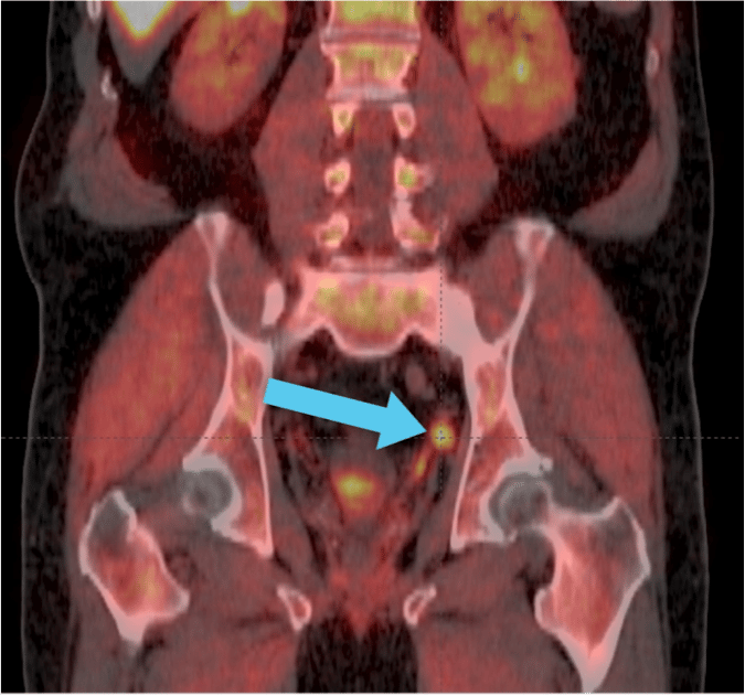
What Is A Pet Scan
A PET scan ) is an imaging medical exam to determine where the disease is in your body.
In the case of cancer, where cells have a high metabolic rate, a radiotracer is injected into the vein or by swallowing where then the diseased cells sort of gobble up the tracers which causes the cancer cells to light up on imaging.
The lit up areas indicates where cancer cells are located, if there is a recurrence after treatment, and if treatment is working.
About Dr Dan Sperling
Dan Sperling, MD, DABR, is a board certified radiologist who is globally recognized as a leader in multiparametric MRI for the detection and diagnosis of a range of disease conditions. As Medical Director of the Sperling Prostate Center, Sperling Medical Group and Sperling Neurosurgery Associates, he and his team are on the leading edge of significant change in medical practice. He is the co-author of the new patient book Redefining Prostate Cancer, and is a contributing author on over 25 published studies. For more information, contact the Sperling Prostate Center.
Search the spc blog
What Radiotracers Are Used In Prostate Cancer
There are numerous radioactive tracers used during a PET scan that helps locate problem areas in the body after prostate cancer.
The most common is a sugar tracer called F-fluoro-2-deoxy-D-glucose as a result of the Warburg effect
Prostate cancer cells vary greatly between slow moving and fast moving cancer cells.
PET Scans using FDG is not useful in detecting primary organ-confined prostate cancer, detecting local recurrence after radical prostatectomy, or in differentiating between post-operative scar and local recurrence for a few reasons.
Prostate cancer is slow growing and may not have a high metabolic rate, which results in low FDG uptake. In addition, FDG-PET cannot reliably differentiate between benign prostate hypertrophy and cancer, and the uptake of the tracer does not correlate with the tumor grade or stage
This glucose tracer is useful in detecting bone and soft-tissue prostate cancer metastases, although it is less sensitive, therefore not as good as a bone scan for prostate cancer
Lastly, FDG tracer are now less favorable in use for prostate cancer compared to others discussed below.
You May Like: Is Fish Oil Bad For Men’s Prostate
How Does Axumin Work
While normal bone scans use radioactive substances that indicate which bones are affected by cancer, Axumin links a radioactive tracer to a certain amino acid that the cancer processes much faster than ordinary cells. This tracer becomes concentrated in cancer, making it visible in bones and soft tissues as well. Salvage Radiation Therapy needs to be initiated at lower PSA levels for best results, and Axumin allows detection at these levels that standard scans do not. An earlier detection combined with a more specific treatment will yield significant benefits to patients with recurrent prostate cancer, who will be able to get a head start on their recovery and better manage their illness.
Our Expert Radiology Services are Offered in Pioneer Valley in Partnership with Baystate Health Locations.
Why Axumin Is So Important

Being able to detect early metastatic disease with a scan offers two important therapeutic advantages. First, the knowledge of where the cancer is located can help guide effective therapy to that specific area of the body and limit damage to other areas of the body. The scan detects where the cancer is not present and where treatment is not needed.
The second valuable contribution that an accurate scan offers is a deeper insight into the disease process itselfrevealing whether or not the cancer has metastasized, and if it has metastasized, to what degree.
Recurrent cancer signaled by a rising PSA is not always due to metastases. Sometimes the cancer remains near or in where the prostate used to be, so PSA is coming from cancer recurring in the prostate gland after radiation or in the prostate fossa after surgery , which is known as a “local recurrence.”
PSA can also be elevated due to growing cancer that has metastasized to the lymph nodes or bones. This is called a systemic recurrence. Systemic recurrences are tremendously more dangerous than local recurrences. Why? A metastasis shows that the cancer has the biologic capacity to spread around the bodya process that ultimately leads to death in more than half of prostate cancer patients. Thus, knowing the location of the recurrence answers an extremely important question: whether the recurrent disease is aggressive enough to metastasize.
Also Check: Can Prostatitis Cause A High Psa
Why Are Pet Scans Important For Prostate Cancer
After definitive treatment for prostate cancer, there can be a recurrence up to 50% of the times but the patient may not know where the recurrence is occurring until the PSA is very high, 20ng/ml or higher. Again, this is after either radiation treatment or surgery. PET scans, especially those with improved radiotracers can detect cancer cells much earlier after prostate cancer treatment.
The Blue Earth Diagnostics Team Offers Reimbursement Support
- Benefit Investigation: Investigation of patient insurance benefits, including deductible and copayments, and verification of coverage status for Axumin and PET/CT, including insurance requirements and possible coverage restrictions.
- Prior Authorization Assistance: Information on PA requirements.*
- Appeal Assistance: Information on appeals requirements for denied Prior Authorizations or denied claims.
You May Like: Can An Ultrasound Detect Prostate Cancer
Interpretation Of Axumin Scans
Interpretation for PET/CT examinations using Axumin are different from standard FDG PET/CT imaging. Axumin specific training is available and is recommended for every interpreting physician. Below is a general guideline for interpretation, taken from Blue Earth Diagnostics Axumin site:
Prostate Bed Focal uptake, visually equal to or greater than bone marrow, in sites typical for prostate cancer recurrence is suspicious for cancer. If focus of uptake is small , it may be considered suspicious if the uptake is visually greater than blood pool.
Prostate Bed Moderate focal asymmetric uptake, visually equal to or greater than bone marrow, is suspicious for cancer recurrence. If focus of uptake is small and in a site typical for recurrence, it may still be considered suspicious if the uptake is visually greater than blood pool.
- Diffuse and heterogeneous uptake visually greater than blood pool is suspicious for cancer.
- Diffuse and homogeneous uptake visually greater than bone marrow is suspicious for cancer.
Lymph Nodes Uptake, visually equal to or greater than bone marrow, is considered suspicious for cancer. If node is small and in a site typical for recurrence, it may still be considered suspicious if visually greater than blood pool. If borderline visually, quantitation using a node to background ratio may help.
Image interpretation is predominantly qualitative but, for areas of borderline increased uptake, an uptake ratio may be supportive.
A Phase Ii Study To Evaluate Axumin Pet/ct For Risk Stratification For Prostate Cancer
| The safety and scientific validity of this study is the responsibility of the study sponsor and investigators. Listing a study does not mean it has been evaluated by the U.S. Federal Government. Read our disclaimer for details. |
| First Posted : December 14, 2017Results First Posted : June 4, 2020Last Update Posted : May 25, 2022 |
DESCRIPTION OF DRUG
Mechanism of action:
Fluciclovine F 18 is a synthetic amino acid transported across mammalian cell membranes by amino acid transporters, such as LAT-1 and ASCT2, which are upregulated in prostate cancer cells. Fluciclovine F 18 is taken up to a greater extent in prostate cancer cells compared with surrounding normal tissues.
Pharmacodynamics:
Following intravenous administration, the tumor-to-normal tissue contrast is highest between 4 and 10 minutes after injection, with a 61% reduction in mean tumor uptake at 90 minutes after injection.
Pharmacokinetics:
Distribution: Following intravenous administration, fluciclovine F 18 distributes to the liver , pancreas , lung , red bone marrow and myocardium . With increasing time, fluciclovine F 18 distributes to skeletal muscle.
TEST PRODUCT, DOSE, AND ROUTE OF ADMINISTRATION All subjects will receive a single IV dose of 10mCi +20%18F-fluciclovine immediately prior to PET scan.
Imaging protocol.
Also Check: Prostate Surgery After Age 70
What Is A Pet Scan And Why Is It Used For Prostate Cancer
New GE Discovery IQ PET Scanner
- What is a PET scan? A PET Scan, short for Positron Emission Tomography Scan, is an imaging technique that uses radioactive tracers to clearly image targeted areas in the body. It is primarily used in the diagnosis, initial staging and treatment strategy, to assess the effectiveness of therapy and for restaging, or the evaluation for recurrent disease after treatment.
- Why is a PET scan used for prostate cancer? PET scanning is used for prostate cancer because of its superior ability to target and capture images of prostate cancer on a cellular level. This allows for more accurate staging and restaging in the overall prostate cancer treatment strategy. With new radiotracers being developed and studied, PET scanning continues to lead the way in what will be possible for imaging during treatment of prostate cancer.
PET scanning has revolutionized the way prostate cancer is imaged because of its ability to target cancer on a cellular and molecular level. PET Scanning uses a radioactive tracer that is absorbed and visualized in cancerous cells related to the prostate. This makes a PET scan a better choice than more conventional imaging modalities that only evaluate an anatomical snapshot of abnormalities rather than the functional aspects of a PET scan.
Axumin Pet Scanning For Prostate Cancer Care
Axumin is an FDA-approved agent used for Axumin PET scans for prostate cancer. Axumin is often able to image and restage recurrent prostate cancer better than any other conventional imaging techniques. Biochemical recurrence, typically suspected with rising PSA levels, is the standard in monitoring patients for suspected recurrent prostate cancer. Traditional imaging techniques are often limited in that they may detect a small lymph node or suspicious finding, but cannot further functionally characterize the molecular activity to determine the level of suspicion. The introduction of Axumin PET scanning has been a breakthrough, allowing physicians the ability to accurately locate and restage prostate cancer with precision, especially in the setting of suspiciously rising PSA levels.
How Do Axumin PET Scans Work?
An Axumin PET uses a radioactive tracer, given as an injection, that is linked to an amino acid which is absorbed by prostate cancer at a much more rapid rate than normal cells. The rapid uptake of Axumin by prostate cancer cells is then imaged by the advanced technology within the PET scan equipment. The PET scan images are then reviewed in order to determine if there has been any spread to other areas in the body.
Recommended Reading: What Does It Mean To Have Aggressive Prostate Cancer
What Are The Possible Side Effects Of Axumin
Most commonly reported adverse reactions are:
- Injection site pain
- Injection site redness
- Unusual taste in the mouth
Tell your doctor if you have any side effect that bothers you or does not go away.
These are not all the possible side effects of Axumin. For more information, ask your doctor or pharmacist. Call your doctor for medical advice about side effects. You may report side effects to FDA at 1-800-FDA-1088.
Axumin Pet Scans: A Breakthrough For Prostate Cancer

Axumin is an FDA-approved, Medicare-covered scan that can achieve early detection of recurrent prostate cancer after surgery or radiation. For years we have been able to detect prostate cancer recurrences with PSA, but standard body and bone scans have been unable to determine the location of the cancer until the PSA level is excessively elevated .
Axumin can detect recurrent disease with PSA levels less than 10 and sometimes much lower, which is the reason this scan is such an important development.
Why Is Axumin So Important?
Being able to detect early metastatic disease with a scan offers two important therapeutic advantages. First, the knowledge of where the cancer is located can help guide effective therapy to that specific area of the body and limit damage to other areas of the body. The scan detects where the cancer is not present and where treatment is not needed.
The second valuable contribution that an accurate scan offers is a deeper insight into the disease process itselfrevealing whether or not the cancer has metastasized, and if it has metastasized, to what degree.
Recurrent cancer signaled by a rising PSA is not always due to metastases. Sometimes the cancer remains near or in where the prostate used to be, so PSA is coming from cancer recurring in the prostate gland after radiation or in the prostate fossa after surgery , which is known as a local recurrence.
How Does Axumin Work?
How Is the New Information Provided by Axumin Utilized?
Also Check: How To Reduce Prostate Enlargement Naturally
Improving Pet Scans Are Good News For Doctors And Patients Alike
- By Charlie Schmidt, Editor, Harvard Medical School Annual Report on Prostate Diseases
A recent blog post discussed a newly approved imaging agent with an unwieldy name: gallium-68 PMA-11. Delivered in small amounts by injection, this minimally radioactive tracer sticks to prostate cancer cells, which subsequently glow and reveal themselves on a positron emission tomography scan. Offered to men with rising PSA levels after initial prostate cancer treatment , this sort of imaging can allow doctors to find and treat new tumors that they might otherwise miss. With currently available imaging technology, such tumors could potentially escape detection until they were larger and more dangerous.
But while gallium-68 PMA-11 is the latest PET tracer to win FDA approval, not everyone can get it. In the United States, its currently available only to patients treated at the University of California, Los Angeles, or the University of California, San Francisco, where the tracer is manufactured. However, two other PET tracers approved for prostate cancer imaging in the US are becoming more accessible.
In January 2021, a team at Stanford University published findings showing that one those tracers, called fluciclovine F18 , identified significantly more metastatic cancers than other conventional types of imaging. Axumin was approved in 2016, and these are among the first data to show how well the tracer performs in real-world settings.
About the Author
What Is An Elevated Psa Level
After your initial prostate cancer treatment, you likely had regular checkups with your doctor. These checkups usually include a blood test to monitor your prostate specific antigen .
If your blood test shows that your PSA has gone up after surgery, radiation, or hormone therapy, your doctor will likely order another PSA test to confirm the results.
If your PSA is still elevated after the test, recurrent prostate cancer is indicated. Imaging tests may then be scheduled to locate where the prostate cancer has returned in your body.
By learning about my recurrent prostate cancer and talking with my urologist, I can be my own best advocate.
Read Also: Does High Psa Always Mean Prostate Cancer
What Is Axumin Pet/ct
Axuminis an FDA-approved diagnostic imaging agent, also known as a tracer, which may help your physician determine if and where your prostate cancer has returned. Like many imaging tracers, Axumin includes a radioactive element which is used to produce images of your body and its internal organs and tissues. Over time and through a natural process, the fluorine 18 will become non-radioactive, and much of it will leave your body in your urine.
What Are The Most Common Types Of Pet Scans For Prostate Cancer
- PSMA PET Scan: Now FDA approved, they are used to locate, stage, and restage prostate cancer. PSMA PET scans have been FDA approved to be used after a diagnosis of prostate cancer in order to stage and determine if cancer has spread to other parts of the body. They have also been approved for use in locating and restaging recurrent prostate cancer for patients with biochemical recurrence.
- Axumin PET Scan: Currentlyin use primarilyfor the purpose of restaging recurrent prostate cancer in those patients with suspected biochemical recurrence.
- FDG PET Scan: May also be used for staging in particular cases of prostate cancer. In some instances, depending on the prostate cancer tumor type, FDG PET scans may be used for the restaging of recurrent prostate cancer as well.
Also Check: What Causes An Enlarged Prostate In A Young Man
Why Are Breakthroughs Occurring More Frequently
The reason for the acceleration in the frequency of breakthroughs is the culmination of extensive basic research leading to a deeper understanding of cellular biology of prostate cancer. More specifically, the specific genetic mutations that cause uncontrolled cellular growth have been elucidated.
Mutated genes are what makes cancer cells different from normal cells. Now that these mutations can be identified, new medications can be designed to compensate for the abnormally functioning genes. Think of how a software patch might be written by a computer programmer to fix a computer glitch.
In prior years, before our arrival at our present-day understanding of cell biology, new medicines were the result of an arduous, trial and error developmental process. A randomly selected chemical would be administered to cancer cells growing in Petri dishes. If the chemical caused the cancer cells to die, it would be administered to animals with cancer. If the cancer regressed and the animal lived, it would be tested in humans. Successful human trials would then lead to FDA approval and the commercial availability of a new treatment.
Unlike the rationally-designed medications of recent times, the way that these medications discovered by trial and error function was often unknown.
Expanded Hours Of Operation
Telephone support is available Monday through Friday, from 9 AM to 8 PM Eastern time.
*Helpline provides information about PA/appeals requirements, and, at the providers option, submits PA forms completed by the provider to the payer.
This document contains factual information and is not intended to be legal or coding advice. Blue Earth Diagnostics does not guarantee coverage or reimbursement for Axumin. The information provided in this document is based upon current, general coding practices. The existence of billing codes does not guarantee coverage and payment. Payer policies vary and may change without notice. It is the providers responsibility to determine and submit accurate information on claims. This includes submitting such as proper codes, modifiers, charges, and invoices for the services that were rendered. The coding on claims should reflect medical necessity and be consistent with the documentation in the patients medical record.
It is essential that hospitals appropriately and accurately determine codes for items and services and apply appropriate charges, even when the payment is bundled. For example, diagnostic radiopharmaceuticals are packaged but still should be coded and billed in order for the cost to be accurately represented in the claims data.
Read Also: How Long Does Erectile Dysfunction Last After Prostate Surgery
