Detecting And Diagnosing Prostate Cancer
Prostate cancer is often detected during the course of a routine prostate exam and/or the PSA blood test, but diagnosing it may require other procedures.
PSA test: PSA is a protein found in prostate cells that helps to keep semen liquified. Most cases of prostate cancer develop in these cells, so an elevated PSA count may be a sign of prostate cancer. However, PSA results are more of an indicator than a firm diagnostic tooltheres not a certain PSA score that means a man has prostate cancer. Instead, there are various ranges that are considered average for different age groups. If the PSA score is elevated for your age, further testing may be recommended.
PSA levels are measured as ng/mL. According to the :
- Men with a PSA level between 4 and 10 have about a 25 percent chance of having prostate cancer.
- Men with a PSA level higher than 10 have more than a 50 percent chance of having prostate cancer.
Not all men with high PSA levels have prostate cancer. High levels may also be caused by a urinary tract infection, prostatitis or benign prostatic hyperplasia, all of which are noncancerous conditions. Conversely, men with a low PSA level may still develop prostate cancer.
PSA tests are not an indication of how aggressive the prostate cancer may be. Many prostate cancers are slow-growing and dont require immediate treatment.
The National Comprehensive Cancer Network suggests these screening guidelines and recommendations for men older than 45:
How Do Doctors Screen For Prostate Cancer
Screening for a disease involves testing for it even if no symptoms or history of the illness are present. Because all men are at risk for prostate cancer, especially older men, getting screened is an important health care step for any man to take. For men not experiencing prostate cancer symptoms, the most common screening method is a prostate-specific antigen blood test.
A PSA is a protein created by cells within the prostate gland. A blood test designed to assess PSA levels measures the concentration of PSA within a patients blood. Although there is no definitive cutoff number, the higher the PSA level in a mans blood, the greater the chances he has prostate cancer.
Typically, men without prostate cancer have a PSA level under 4 nanograms of PSA per milliliter of blood , which means those with PSA test results above 4 ng/mL may require further testing. While the likelihood of a patient having prostate cancer decreases dramatically with a 4 ng/mL result, some doctors may request that those with lower PSA levels get additional testing. When done in tandem with other scans and tests, a PSA blood test can help medical professionals provide a more accurate prostate cancer diagnosis.
Getting A Prostate Ultrasound For Prostate Cancer
A prostate ultrasound is often used early as a way of diagnosing prostate cancer. Prostate cancer develops in the prostate, a small gland that makes seminal fluid and is one of the most common types of cancer in men.
Prostate cancer usually grows over time, staying within the prostate gland at first, where it may not cause serious harm. While some types of prostate cancer grow slowly and may need minimal or no treatment, other types are aggressive and can spread quickly. The earlier you catch your prostate cancer, the better your chance of successful treatment.
If your doctor suspects you might have prostate cancer they will conduct a number of tests which may include a prostate-specific antigen test, a digital exam of your prostate, and an ultrasound. If your blood work comes back and your PSA is high, your prostate feels abnormal upon exam and the ultrasound show signs of cancer, your doctor will likely want to do a biopsy.
What Does It Show
An ultrasound machine creates images called sonograms by giving off high-frequency sound waves that go through your body. As the sound waves bounce off organs and tissues, they create echoes. The machine makes these echoes into real-time pictures that show organ structure and movement and even blood flow through blood vessels. The images can be seen on a computer screen.
Ultrasound is very good at getting pictures of some soft tissue diseases that dont show up well on x-rays. Ultrasound is also a good way to tell fluid-filled cysts from solid tumors because they make very different echo patterns. Its useful in some situations because it can usually be done quickly and doesnt expose people to radiation.
Ultrasound images are not as detailed as those from or scans. Ultrasound cannot tell whether a tumor is cancer. Its use is also limited in some parts of the body because the sound waves cant go through air or through bone.
Doctors often use ultrasound to guide a needle to do a biopsy . The doctor looks at the ultrasound screen while moving the needle and can see the needle moving toward and into the tumor.
For some types of ultrasound exams, the transducer is pushed against and moved over the skin surface. The sound waves pass through the skin and reach the organs underneath. In other cases, to get the best images, the doctor must use a transducer thats put into a body opening, such as the esophagus , rectum, or vagina.
Tests To Diagnose And Stage Prostate Cancer

Most prostate cancers are first found as a result of screening. Early prostate cancers usually dont cause symptoms, but more advanced cancers are sometimes first found because of symptoms they cause.
If prostate cancer is suspected based on results of screening tests or symptoms, tests will be needed to be sure. If youre seeing your primary care doctor, you might be referred to a urologist, a doctor who treats cancers of the genital and urinary tract, including the prostate.
The actual diagnosis of prostate cancer can only be made with a prostate biopsy .
On this page
Where To Find The Ultrasound Equipment You Need
Is your practice ready to provide cancer screenings for men? Testicular ultrasounds, prostate ultrasounds and prostate biopsies are universally agreed upon as advanced diagnostic tools. If you want to learn even more about these ultrasound tools, . Be prepared for mens health screenings this month and find the ultrasound equipment thats right for your practice!
Importance Of A Prostate Ultrasound For Early Detection
Men need to realize the importance of early detection. This is especially the case when it comes to the diagnosis of prostate cancer. Several tests can assist in the early detection of prostate cancer. A rectal exam is often what first identifies an abnormality with the prostate gland.
Following this type of finding, PSA testing will often be advised. At this point, levels of PSA in the blood are identified through a special blood test. A Gleason score test may also be requested.
A biopsy is often advised. An ultrasound-guided biopsy is preferred recently, but a standard prostate ultrasound may be utilized first. Even though some men may find the procedure somewhat uncomfortable, it is crucial to understand the importance of the test.
Getting A Prostate Ultrasound
Ultrasound imaging uses high-frequency sound waves to create a picture of the prostate. Ultrasounds are used early in the diagnostic process to determine whether your prostate is enlarged or has an abnormal or asymmetrical shape. If your prostate is enlarged without any other characteristics of cancer, you may just have benign prostate hyperplasia. As you age, your prostate increases in size. An ultrasound can help your doctor determine if your prostate size increase is normal and age-related or a sign of prostate cancer.
Ultrasound is also used very frequently during a prostate biopsy to guide the physician to biopsy exactly where needed. In order to produce an image of the prostate, a thin ultrasound probe is inserted a short distance into the rectum. This probe emits high-frequency sound waves and detects their return. These sound waves can then be detected and measured as they reflect off of various structures inside the body.
When sound waves echo off of an object, they change slightly. The ultrasound machine can interpret these very tiny changes in the character of the returning sound wave to make determinations about the object that it has hit. Different types of structures reflect or echo sound waves differently. These differences can be detected and an image produced that shows where one structure stops and another starts. This allows for a detailed view of the area near the ultrasound probe.
How Do I Get Ready For A Prostate/rectal Ultrasound
-
Your healthcare provider will explain the procedure and you can ask questions. Make a list of questions and any concerns with your healthcare provider before the procedure. Consider bringing a family member or trusted friend to the medical appointment to help you remember your questions and concerns.
-
You may be asked to sign a consent form that gives your permission to do the procedure. Read the form carefully and ask questions if something is not clear.
-
You may be asked to stop taking blood-thinning medicines, such as aspirin, for a week or so before the test if it is being done as part of a biopsy.
-
You usually do not need to stop eating or drinking before the test. You also usually will not need medicine to help you relax .
-
You may be given a small enema before the test.
-
Follow any other instructions your provider gives you to get ready.
Changing Standard Of Care
Will a Prostate MRI eventually become the standard of care? Dr. Walker thinks so. Across the country and in the minds of urologists, he says the Prostate MRI is becoming essential for the management of patients at risk of prostate cancer.
Now I think a lot of docs are becoming more comfortable using the MRI ahead of time to get a little bit more accurate biopsy. And more patients actually know about the MRI technology and even ask about it. -Dr. Todd Brandt of Metro Urology
How Do I Prepare For A Prostate Ultrasound
Some possible instructions that your doctor might give you before the test include:
- Dont eat for a few hours before the test.
- Take a laxative or enema to help clear out your intestines a few hours before the test.
- Stop taking any medications that can thin your blood, such as nonsteroidal anti-inflammatory drugs or aspirin, about a week before the procedure. This is usually recommended if your doctor plans to take a biopsy of your prostate.
- Dont wear any jewelry or tight clothes to the clinic on the day of the procedure.
- Take any medications recommended to help you relax during the procedure. Your doctor may recommend a sedative, such as lorazepam .
- Make sure someones available to take you home in case your doctor gives you a sedative.
How Is The Procedure Performed
In men, the prostate gland is located directly in front of the rectum, so the ultrasound exam is performed transrectally in order to position the imaging probe as close to the prostate gland as possible.
For a transrectal ultrasound, you will be asked to lie on your side with your knees bent. A disposable protective cover is placed over the transducer, it is lubricated, inserted through the anus and placed into the rectum.
The images are obtained from different angles to get the best view of the prostate gland.
If a suspicious lesion is identified with ultrasound or with a rectal examination, an ultrasound-guided biopsy can be performed. This procedure involves advancing a needle into the prostate gland while the radiologist watches the needle placement with ultrasound. A small amount of tissue is taken for microscopic examination.
A prostate-specific antigen test, which measures the amount of PSA in the blood, may be administered to determine if a patient is at high risk for cancer. In this case, a biopsy is performed and an ultrasound probe is used to guide the biopsy to specific regions of the prostate gland.
When the exam is complete, you may be asked to dress and wait while the ultrasound images are reviewed.
This ultrasound examination is usually completed in less than 20 minutes.
When To Consider A Prostate Mri

Studies indicate that MRI may be helpful in the following situations. The best images are obtained when using an endorectal coil.
- You have a PSA that continues to increase, but an ultrasound-guided prostate biopsy does not reveal cancer; an MRI may be able to better pinpoint a suspicious area for a more targeted biopsy and increase the likelihood of finding cancer if it is there.
- Different elements of your diagnostic workup are in conflict ; an MRI can better determine size of the tumor and whether it has extended beyond the capsule.
- For large palpable tumors, MRI can rule out cancer that extends beyond the prostate itself.
- If your PSA rises following prostate cancer treatment, MRI can be used to identify any cancerous tissue in the periprostatic bed , which indicates a local recurrence.
- MRI may provide better guidance about where to target radiation therapy.
What Happens During A Prostate/rectal Ultrasound
You may have a prostate/rectal ultrasound done as an outpatient or during ahospital stay. The way the test is done may vary depending on yourcondition and your healthcare provider’s practices.
Generally, a prostate/rectal ultrasound follows this process:
You will need to remove any clothing, jewelry, or other objects that may get in the way of the procedure.
If asked to remove clothing, you will be given a gown to wear.
You will lie on an exam table on your left side with your knees bent up to your chest.
The healthcare provider may do a digital rectal exam before the ultrasound.
The provider puts a clear gel on the transducer and puts the probe into the rectum. You may feel a fullness of the rectum at this time.
The provider will turn the transducer slightly several times to see different parts of the prostate gland and other structures.
If blood flow is being looked at, you may hear a whoosh, whoosh sound when the Doppler probe is used.
Once the test is done, the provider will wipe off the gel.
A prostate/rectal ultrasound may be uncomfortable and you will need toremain still during the test. The gel will also feel cool and wet. Thetechnologist will use all possible comfort measures and do the scan asquickly as possible to minimize any discomfort.
Other Tests To Detect Cancer
Can ultrasound detect cancer? It can detect changes that appear to be cancerous. If an ultrasound detects abnormalities that look like cancer changes in tissues, more tests will be needed to confirm the diagnosis. The following tests may be used to get a better look at things:
- CT Scan
- Biopsy
- Tumor Marker Tests
- Surgical Intervention
How Accurate Is A Prostate Ultrasound
A prostate ultrasound is more accurate than an X-ray. This is because your technician can see the images as the transducer moves through your rectum rather than having to take a snapshot and develop the images. Ultrasound tests are also safer than X-rays because they dont produce any dangerous radiation.
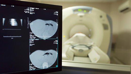
A prostate ultrasound is also faster than a computed tomography test, which provides 3-D images of your prostate and the areas around it. CT scans require more preparation and time for testing, and they dont provide real-time images.
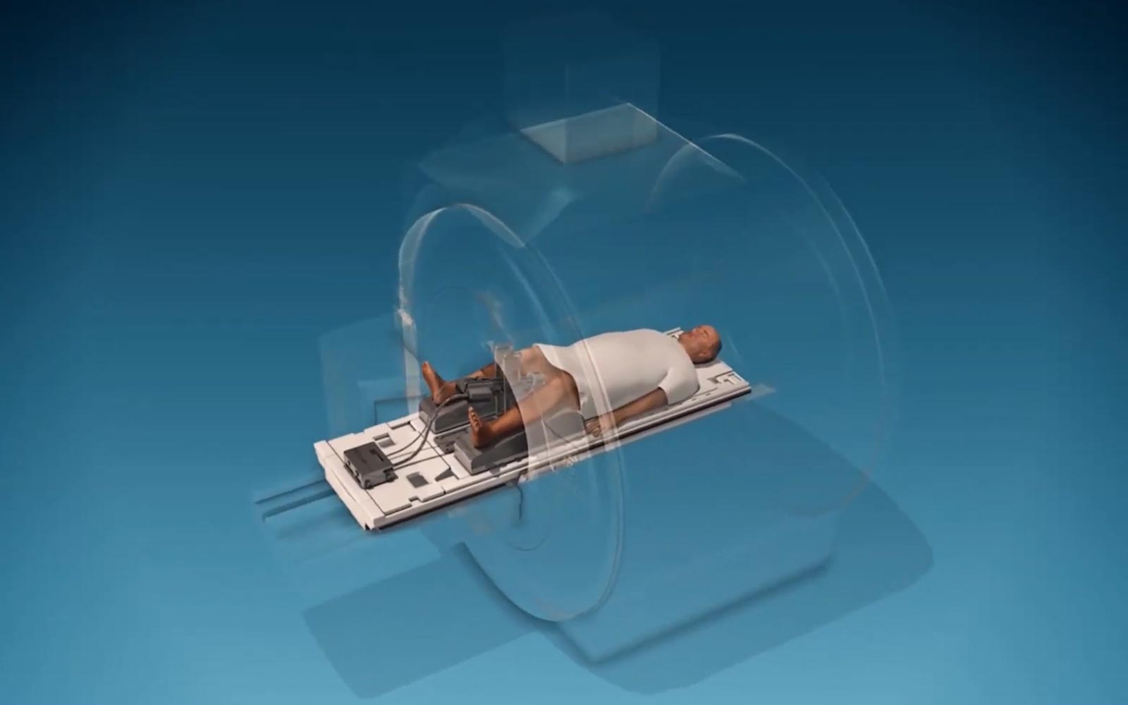
Will The Mri Be Done With An Endorectal Coil Or An External Pelvic Coil
Some radiology practices use an endorectal coil a probe-like device covered with latex which is inserted into the rectum and helps provide high-quality images of the prostate. With a newer, high-quality MRI system, endorectal coils are not necessary and an external pelvic coil can be used instead, eliminating patient discomfort while maintaining high quality images.
What Kind Of Images Does Trus Take
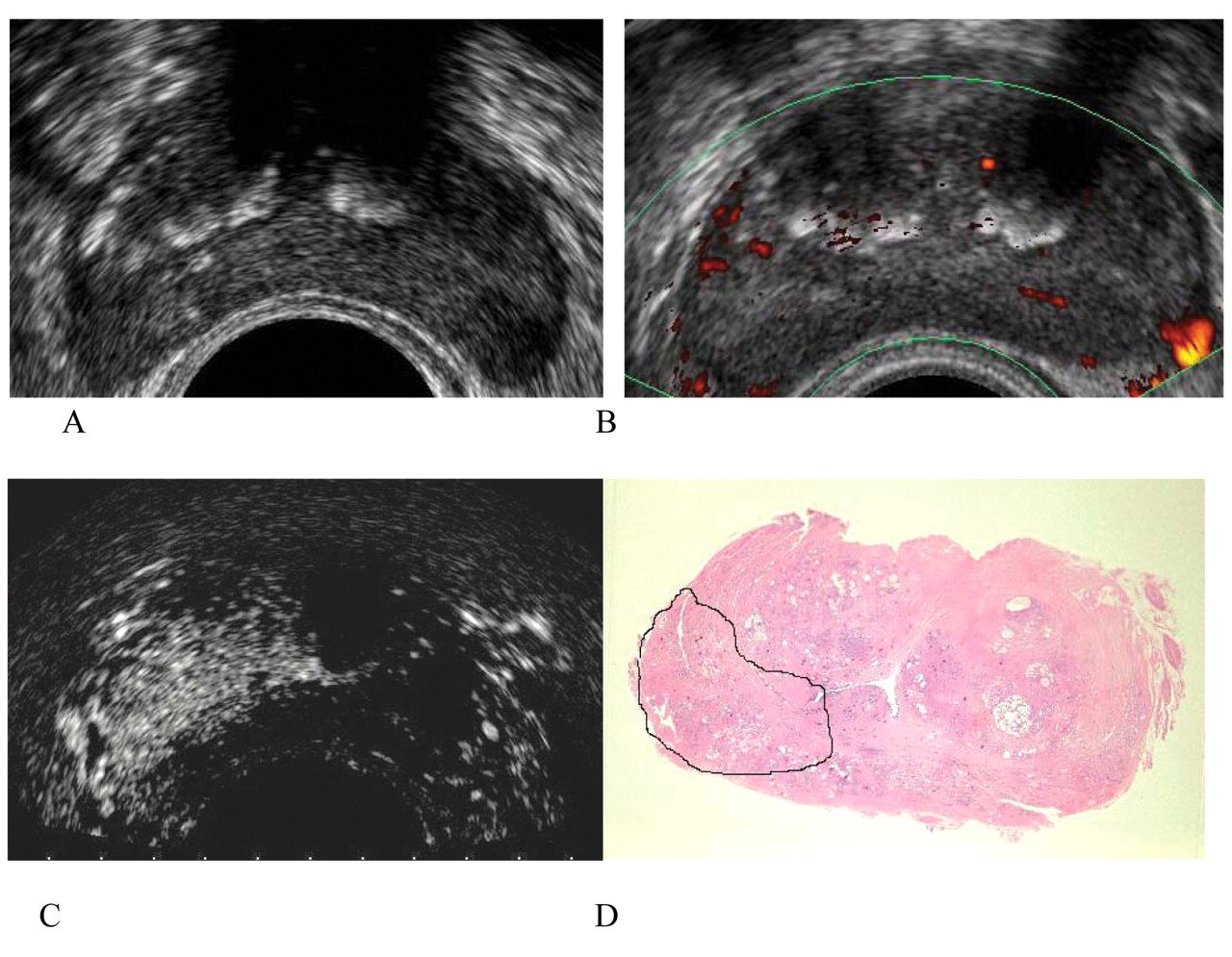
The rectal probe sends sound waves to the prostate gland; normal and abnormal tissue bounce back different kinds of echoes that are relayed to the computer, which translates their pattern into a video picture of the prostate.
- Isoechoic areas represent normal tissue and bounce back, or echo, the same amount sound waves as they received.
- Hypoechoic areas send back significantly fewer echoes than they received and often indicate the presence of cancer.
- Hyperechoic areas send back significantly more echoes than they received and often indicate the presence of prostatic calcifications, or tiny stones, in the prostate. The stones are usually harmless unless they become infected.
Positron Emission Tomography Scan
A PET scan is similar to a bone scan, in that a slightly radioactive substance is injected into the blood, which can then be detected with a special camera. But PET scans use different tracers that collect mainly in cancer cells. The most common tracer for standard PET scans is FDG, which is a type of sugar. Unfortunately, this type of PET scan isnt very useful in finding prostate cancer cells in the body.
However, newer tracers, such as fluciclovine F18, sodium fluoride F18, and choline C11, have been found to be better at detecting prostate cancer cells.
Other newer tracers, such as Ga 68 PSMA-11 and 18F-DCFPyl , attach to prostate-specific membrane antigen , a protein that is often found in large amounts on prostate cancer cells. Tests using these types of tracers are sometimes referred to as PSMA PET scans.
These newer types of PET scans are most often used if its not clear if prostate cancer has spread. For example, one of these tests might be done if the results of a bone scan arent clear, or if a man has a rising PSA level after initial treatment but its not clear where the cancer is in the body.
The pictures from a PET scan arent as detailed as MRI or CT scan images, but they can often show areas of cancer anywhere in the body. Some machines can do a PET scan and either an MRI or a CT scan at the same time, which can give more detail about areas that show up on the PET scan.
How To Prepare For A Prostate Ultrasound
Ultrasound imaging of prostate tissue is a relatively simple procedure. Still, men should know how they can best prepare for the entire process. This helps to make the man more comfortable while they are undergoing the procedure.
There are not too many steps that a man can take to prepare for the procedure. There are, however, a few things that a man can do to make the entire procedure less inconvenient for themselves.
What Is A Trus

Ultrasound scans use high frequency sound waves to create a picture of a part of the body. A prostate ultrasound scan can show up changes in your prostate, including abnormal growths.
The ultrasound scanner has a microphone that gives off sound waves. The sound waves bounce off the organs inside your body, and the microphone picks them up. The microphone links to a computer. This turns the sound waves into a picture on the screen.
You might have a sample of tissue taken from the prostate gland at the same time. This is called transrectal ultrasound guided biopsy.
Enhanced Ultrasound Improves Detection Of Prostate Cancer
According to a recent article published in The Journal of Urology, the use of contrast enhanced color Doppler in endorectal ultrasound improves the detection of prostate cancer.
Prostate cancer is a common cancer among men in the United States. Prostate cancer is the second leading cause of cancer death in men in the United States. The prostate is a walnut-size gland that is located between the bladder and rectum and forms a component of semen. Prostate specific antigen levels , a digital rectal exam and transrectal ultrasound are common tests used to detect prostate cancer. If any suspicious mass is found through these tests, a patient must then undergo biopsies to definitively determine whether cancer exists. However, it is imperative that a physician takes a biopsy from the area in the prostate where the cancer exists to provide accurate diagnostic information. Physicians often use endorectal ultrasound to help determine where in the prostate to take a biopsy. Researchers are attempting to improve upon the accuracy of ultrasound in the guidance of placement of biopsies, including the introduction of contrast, which help physicians to discern between healthy looking tissue and possible sites of cancer.
Reference: Roy C, Buy X, Lang H, et al. Contrast enhances color Doppler endorectal sonography of the prostate: efficiency for detecting peripheral zone tumors and role for biopsy procedure.
The Journal of Urology. 2003;170:69-72.
Has The Radiologist Who Will Read The Study Undergone Pi
The Prostate Imaging Reporting and Data System, or PI-RADS®, refers to standards that have been developed by several organizations to improve early diagnosis and treatment of prostate cancer.
Prostate cancer is the most commonly diagnosed cancer in men, with one out of nine men diagnosed in their lifetime. And while many cases dont require treatment, it is a leading cause of cancer death among men, second only to lung cancer. The broad range between cancer that does not require treatment and life-threatening prostate cancer means that proper diagnosis is key to determining next steps.
Transrectal Ultrasound Guided Biopsy
This test uses sound waves produced by a small probe placed in the rectum to create an image of the prostate on a video screen. The echoes from the waves are translated by a computer into a picture, which can show the location of more suspicious or abnormal areas of the prostate.
Since cancer, even if present, may not be seen with the TRUS, it is important to sample the entire prostate gland. An instrument called a biopsy gun quickly inserts and removes narrow needles, obtaining small cores of tissue that are sent to the laboratory for examination. From six to 18 cores may be removed from different areas of the prostate, especially from the more suspicious locations.
The procedure takes less than half an hour, and usually causes only a little discomfort and occasionally some bleeding. An antibiotic such as Ciproflaxacin is usually given before and after the procedure to reduce risk of infection.
Sometimes, the first biopsy doesn’t reveal the presence of cancer even when cancer is strongly suggested by the patient’s symptoms or PSA test results. Repeat biopsies may be required before the cancer is actually discovered.
What Are The Limitations Of Prostate Ultrasound Imaging
Men who have had the tail end of their bowel removed during prior surgery are not good candidates for ultrasound of the prostate gland because this type of ultrasound typically requires placing a probe into the rectum. However, the radiologist may attempt to examine the prostate gland by placing a regular ultrasound imaging probe on the perineal skin of the patient, between the legs and behind the scrotum of the patient. Sometimes the gland can be examined by ultrasound this way, but the images may not be as detailed as with the transrectal probe. An MRI of the pelvis may be obtained as an alternative imaging test, because it may be obtained with an external receiver coil.
Biopsy During Surgery To Treat Prostate Cancer
If there is more than a very small chance that the cancer might have spread , the surgeon may remove lymph nodes in the pelvis during the same operation as the removal of the prostate, which is known as a radical prostatectomy .
The lymph nodes and the prostate are then sent to the lab to be looked at. The lab results are usually available several days after surgery.
What Is A Prostate/rectal Ultrasound
A prostate or rectal ultrasound is an imaging test that uses sound waves tolook at your prostate or your rectum.
The healthcare provider uses a small probe called a transducer to make theimages of your prostate or rectum. The transducer is about the size of afinger. It is gently placed into your rectum, where it sends out soundwaves that bounce off your organs and other structures. The sound waves aretoo high-pitched for you to hear. The transducer then picks up the bouncedsound waves. These are made into pictures of your organs.
Your provider can add another device called a Doppler probe to thetransducer. This probe lets your provider hear the sound waves thetransducer sends out. He or she can hear how fast blood is flowing througha blood vessel and in which direction it is flowing. No sound or a faintsound may mean that you have a blockage in the flow.
