How Does The Procedure Work
Ultrasound imaging is based on the same principles involved in the sonar used by bats, ships and fishermen. When a sound wave strikes an object, it bounces back, or echoes. By measuring these echo waves, it is possible to determine how far away the object is as well as the object’s size, shape and consistency. This includes whether the object is solid or filled with fluid.
In medicine, ultrasound is used to detect changes in the appearance of organs, tissues, and vessels and to detect abnormal masses, such as tumors.
In an ultrasound exam, a transducer both sends the sound waves and records the echoing waves. When the transducer is pressed against the skin, it sends small pulses of inaudible, high-frequency sound waves into the body. As the sound waves bounce off internal organs, fluids and tissues, the sensitive receiver in the transducer records tiny changes in the sound’s pitch and direction. These signature waves are instantly measured and displayed by a computer, which in turn creates a real-time picture on the monitor. One or more frames of the moving pictures are typically captured as still images. Short video loops of the images may also be saved.
The same principles apply to ultrasound procedures such as transrectal ultrasound which require insertion of a special imaging probe or transducer into the body.
How Soon Will Prostate Test Results Be Available
Results for simple medical tests such as some urodynamic tests, cystoscopy, and abdominal ultrasound are often available soon after the test. The results of other medical tests such as PSA blood test and prostate tissue biopsy may take several days to come back. A health care provider will talk with the patient about the results and possible treatments for the problem.
What Is The Function Of The Prostate
The prostate plays a fundamental role in the formation of seminal fluid, making sure it produces the correct volume. Their secretions also benefit the mobility of sperm, ensuring that they arrive safely to the egg to fertilize it.
Additionally, this gland is responsible for protecting men from urinary tract infections and maintains a healthy bladder, so it is paramount that it is in perfect condition.
Also Check: What Happens If You Have Prostate Cancer
Prostate Enlargement And Psa
Cells of the prostate gland release protein known as Prostate Specific Antigen into the bloodstream. This protein can circulate in the bloodstream in two ways: on its own or bound to other proteins. PSA tests are usually conducted to measure the quantity of this antigen in the blood. While free-PSA test measures the quantity of unbound PSA and bound-PSA test measures the quantity of bound PSA, total PSA test in general measures the sum total of both free and bound PSA in the blood.
For Confidence On 2 Or

We know that sample variance s2 drawn randomly from a normal population is distributed as 2×2/df. A confidence-type statement on 2 from this relationship, with the additional twist that the asymmetric distribution requires the chi-square values excluding 1/2 in each tail to be found separately, is given by
R.H. Riffenburgh, in, 2012
Recommended Reading: Viagra And Frequent Urination
Comparison Of Measured Prostate Volume And Trus Estimated Prostate Volume
We further examined the percent-difference in volumes between measured and estimated volumes using the three formulas for our primary cohort . In comparison to the measured volume, the ellipsoid formula revealed an underestimation of the volume by a mean of 8.80cc , whereas the bullet formula overestimated prostate volumes by 1.97cc . Finally, when comparing measured prostate volumes to TRUS-estimated volumes using our new formula with 0.66 as the coefficient, there was an overestimation by a mean of 2.76cc .
Renal Doppler Criteria For Renal Artery Stenosis In Native Kidneys
- Direct criteria by evaluating the main renal artery
- Peak systolic velocity > 200 cm/sec. This has the best accuracy for the diagnosis and grading of RAS.
- Renal Aortic Ratio > 3.5. This is the ratio of the PSV in the stenosed renal artery to that PSV in the pre-renal abdominal aorta.
Recommended Reading: Cialis For Prostatitis
How Do I Prepare For A Prostate Ultrasound
Some possible instructions that your doctor might give you before the test include:
- Dont eat for a few hours before the test.
- Take a laxative or enema to help clear out your intestines a few hours before the test.
- Stop taking any medications that can thin your blood, such as nonsteroidal anti-inflammatory drugs or aspirin, about a week before the procedure. This is usually recommended if your doctor plans to take a biopsy of your prostate.
- Dont wear any jewelry or tight clothes to the clinic on the day of the procedure.
- Take any medications recommended to help you relax during the procedure. Your doctor may recommend a sedative, such as lorazepam .
- Make sure someones available to take you home in case your doctor gives you a sedative.
If You Have Any Problems
If you have any discomfort when urinating, any pain in the area around the prostate or any indication of a problem, the recommendation is to visit a urologist who can examine you to determine the source of discomfort or the problem.
It is important to visit a urologist whenever you feel the smallest discomfort, as the problem could get worse with time.
This article is merely informative, oneHOWTO does not have the authority to prescribe any medical treatments or create a diagnosis. We invite you to visit your doctor if you have any type of condition or pain.
If you want to read similar articles to What is the Normal Size of the Prostate, we recommend you visit our Family health category.
You May Like: Does An Enlarged Prostate Affect A Man Sexually
Acute And Chronic Prostatitis
The prevalence of prostatitis ranges between 5% and 11%. Prostatitis occurs at any age and its incidence increases with age. Acute bacterial prostatitis often begins with chills and fever, lower abdominal discomfort, perineal pain and burning on urination. In chronic bacterial prostatitis perineal pain and increased frequency of painful voiding are the most common symptoms. Acute or chronic bacterial prostatitis with confirmed or suspected infection should be distinguished from chronic pelvic pain syndrome , according to the classification suggested by the National Institute of Diabetes and Digestive and Kidney Diseases . The pathophysiology of prostatitis is not well understood. In patients with prostatitis, the activities of prostatic antibacterial factor are decreased and the pH is very alkaline. Bacteria invade the prostate by an ascending urethral infection, by reflux of infected urine into prostatic ducts or by lymphatic/haematogenous spread. Acute bacterial prostatitis appears in US as a hypoechoic rim around the prostate and colour Doppler shows an increased flow . A prostate abscess appears sonographically as a hypoechogenic walled-off collection of fluid. In chronic bacterial prostatitis a diffuse increased enhancement of contrast agent may be found. US contrast agents show an increased perfusion of the prostate during acute and chronic infection, however they are not used in routine clinical practice since no studies regarding this issue have been performed.
What Are The Limitations Of Prostate Ultrasound Imaging
Men who have had the tail end of their bowel removed during prior surgery are not good candidates for ultrasound of the prostate gland because this type of ultrasound typically requires placing a probe into the rectum. However, the radiologist may attempt to examine the prostate gland by placing a regular ultrasound imaging probe on the perineal skin of the patient, between the legs and behind the scrotum of the patient. Sometimes the gland can be examined by ultrasound this way, but the images may not be as detailed as with the transrectal probe. An MRI of the pelvis may be obtained as an alternative imaging test, because it may be obtained with an external receiver coil.
Don’t Miss: Expressed Prostatic Secretions Procedure
Relationship Between Measured And Estimated Prostate Volumes
The estimated volume from TRUS imaging assumes an ellipsoid geometrical shape of the prostate using the formula . In order to identify the best coefficient for this series of 153 fresh prostate specimens, we used the measured prostate weight converted to measured prostate volume by using 1.02g/cc as the density, as defined above. Thus, the mean measured volume of our cohort was 48.1cc .
Measured prostate volumes along with TRUS-obtained prostate dimensions were then used to calculate a new coefficient from the rearranged algebraic formula: Coefficient=V/L x H x W, where L, H and W were all obtained by TRUS, and V is the measured prostatic volume of fresh prostates, obtained just after surgery, as mentioned above. This calculation was performed for each of the 153 prostates, which led to a calculated mean coefficient of 0.66.
Linear regression plots were created in order to compare the newfound coefficient of 0.66 with the ellipsoid coefficient of 0.52. Figure a shows that plotting estimated prostate volumes against measured prostate volumes using 0.66 as a coefficient yielded an equation of y=0.892x+8.8829 with an R2 value of 0.42 . By performing the same analysis using 0.52 as the coefficient, the equation generated was y=0.5652x+13.028 with R2=0.32 .
Fig. 1Fig. 2Fig. 3
What Gets Stored In A Cookie

This site stores nothing other than an automatically generated session ID in the cookie no other information is captured.
In general, only the information that you provide, or the choices you make while visiting a web site, can be stored in a cookie. For example, the site cannot determine your email name unless you choose to type it. Allowing a website to create a cookie does not give that or any other site access to the rest of your computer, and only the site that created the cookie can read it.
Read Also: Do Females Have Prostate Cancer
Why A Psa Test Is Done
A PSA test may be done to:
- help find prostate cancer early in men who dont have any signs or symptoms of the disease
- check for cancer in men who have signs or symptoms of prostate cancer
- confirm a diagnosis when other tests suggest prostate cancer
- predict a prognosis for prostate cancer
- predict if cancer has spread outside the prostate
- plan treatment for prostate cancer
- monitor men with prostate cancer who are being treated with active surveillance
- find out if cancer treatments are working
- find out if cancer has come back after treatment
A PSA test is often used together with a digital rectal exam to increase the chance of finding prostate cancer early when it is easier to treat. Using these tests together is better than using either test alone.
Setting Your Browser To Accept Cookies
There are many reasons why a cookie could not be set correctly. Below are the most common reasons:
- You have cookies disabled in your browser. You need to reset your browser to accept cookies or to ask you if you want to accept cookies.
- Your browser asks you whether you want to accept cookies and you declined. To accept cookies from this site, use the Back button and accept the cookie.
- Your browser does not support cookies. Try a different browser if you suspect this.
- The date on your computer is in the past. If your computer’s clock shows a date before 1 Jan 1970, the browser will automatically forget the cookie. To fix this, set the correct time and date on your computer.
- You have installed an application that monitors or blocks cookies from being set. You must disable the application while logging in or check with your system administrator.
Recommended Reading: Tamsulosin Ejaculation
Bladder Ultrasound Longitudinal View
- Place the transducer with the indicator pointing towards the patients head in the patients midline, right above the pubic symphysis.
- Rock the probe so that it points down towards the pelvic cavity.
POCUS 101 Tip: One of the most important things to remember when performing bladder ultrasound is that the bladder is directly posterior to the pubic bone/symphysis. If you are unable to get proper images, most likely your ultrasound probe is placed too superiorly.
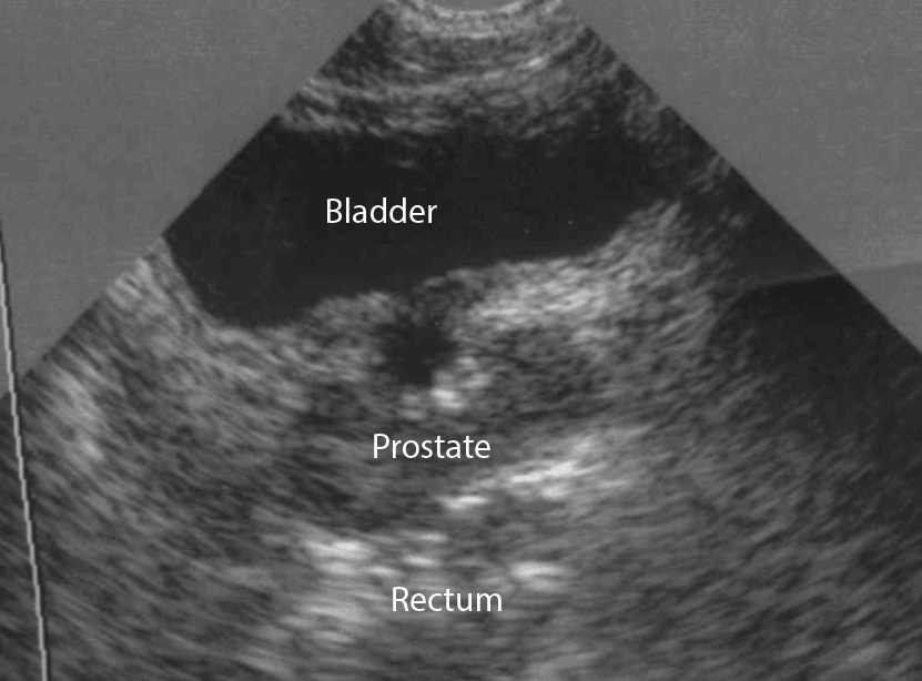
- In the longitudinal view, identify the Bladder, Bowel Gas, Uterus , Prostate , and Rectum.
- Observe the lateral borders of the bladder by tilting/fanning the probe left and right.
Bladder Ureteral Jets For Kidney Stones
Ureteral jets are a normal and periodic efflux of urine from the ureter into the bladder. Visualization of bilateral ureteral jets rules out complete obstruction of a specific ureter with high specificity .
- To see ureteral jets, scan slowly through the bladder in the transverse view and focus on the trigone .
- Turn oncolor Doppler or powerDopplerwhile scanning the bladder in the transverse view. It may take 5-10 minutes before you can visualize bilateral jets so be patient.
Recommended Reading: Does Enlarged Prostate Affect Ejaculation
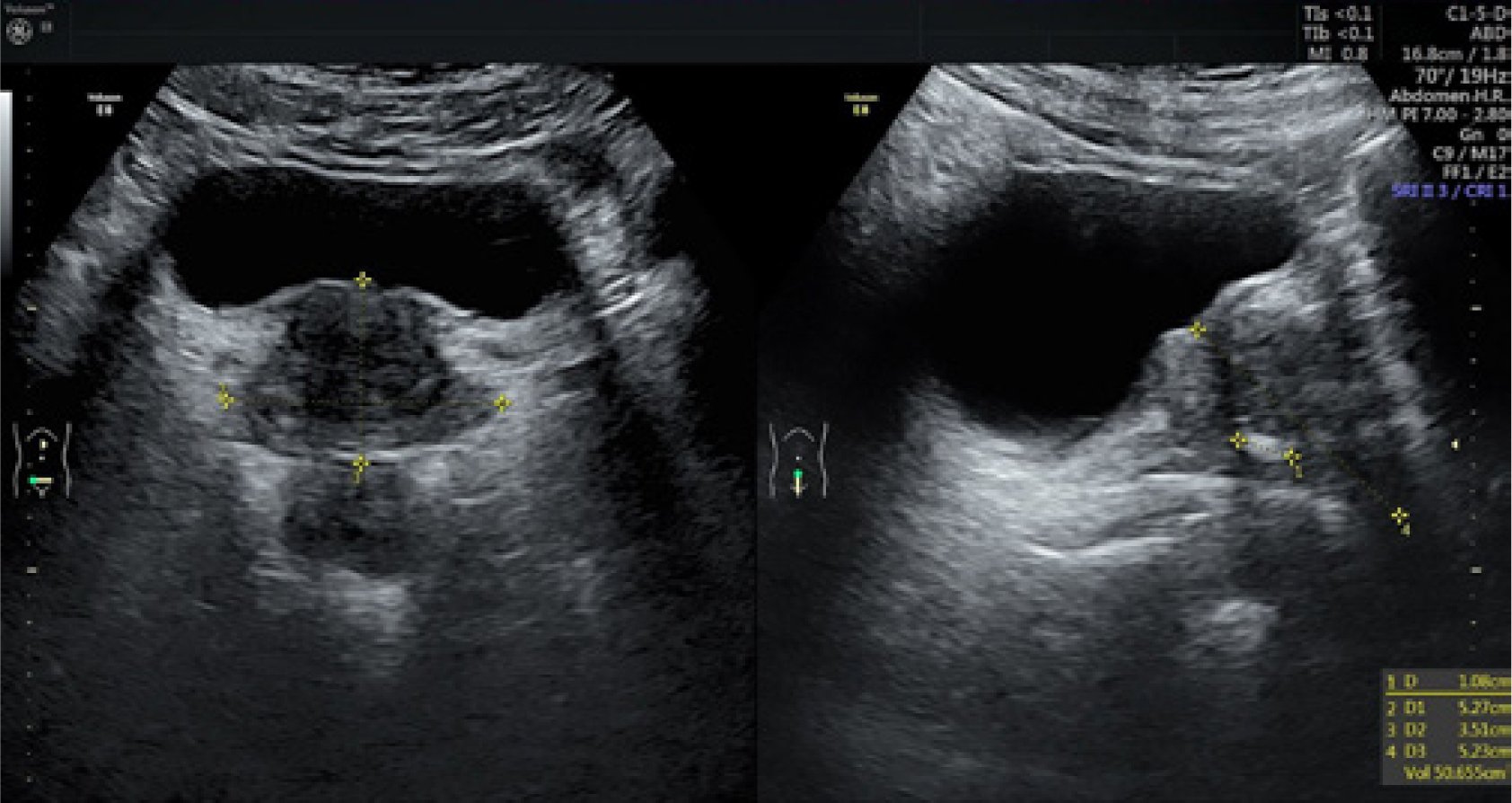
Prostatic Volume Determination By Transabdominal Ultrasonography: Does Accuracy Vary Significantly With Urinary Bladder Volumes Between 50 To 400 Ml
University of Ghana School of Medicine and Dentistry, Korle Bu, Accra, Ghana
Correspondence
Edmund K. Brakohiapa, University of Ghana School of Medicine and Dentistry, Korle Bu, Accra, Ghana. Tel: +23 324 436 7086 E-mail:
University of Ghana School of Medicine and Dentistry, Korle Bu, Accra, Ghana
Correspondence
Edmund K. Brakohiapa, University of Ghana School of Medicine and Dentistry, Korle Bu, Accra, Ghana. Tel: +23 324 436 7086 E-mail:
Example: Distribution Of Prostate Volumes
Figure 5.6 shows prostate volume for 291 patients in the 50- to 89-year age range separated into decades of age: 50s, 60s, 70s, and 80s. The means are shown by solid circles. The whiskers indicate about 2 standard errors above and below the mean, which includes 95% of the data on an idealized distribution. We can see by inspection that the mean volumes appear to increase somewhat by age, but that there is so much overlap in the variability that we are not sure this increase is a dependably real phenomenon from decade to decade. However, we would take a small risk in being wrong by concluding that the increase from the youngest to the oldest is a real change.
Figure 5.6. Prostate volume means for 297 men allocated to their decades of age, with attached whiskers representing about two standard deviations above and below the respective means.
R.H. Riffenburgh, in, 2012
Also Check: Pentostatin Side Effects
What Are Clinical Trials And Are They Right For You
Clinical trials are part of clinical research and at the heart of all medical advances. Clinical trials look at new ways to prevent, detect, or treat disease. Researchers also use clinical trials to look at other aspects of care, such as improving the quality of life for people with chronic illnesses. Find out if clinical trials are right for you.
What Is Ultrasound Imaging Of The Prostate

Ultrasound is safe and painless. It produces pictures of the inside of the body using sound waves. Ultrasound imaging is also called ultrasound scanning or sonography. It uses a small probe called a transducer and gel placed directly on the skin. High-frequency sound waves travel from the probe through the gel into the body. The probe collects the sounds that bounce back. A computer uses those sound waves to create an image. Ultrasound exams do not use radiation . Because images are captured in real-time, they can show the structure and movement of the body’s internal organs. They can also show blood flowing through blood vessels.
Ultrasound imaging is a noninvasive medical test that helps physicians diagnose and treat medical conditions.
Prostate ultrasound, also called transrectal ultrasound, provides images of a man’s prostate gland and surrounding tissue. The exam typically requires insertion of an ultrasound probe into the rectum of the patient. The probe sends and receives sound waves through the wall of the rectum into the prostate gland which is situated right in front of the rectum.
You May Like: Finding The Prostate Externally
Inaccurate Bladder Scan Recordings
There are several reasons why a bladder scan may be inaccurate :
- Part of the bladder extends outside of the scanned region or due to altered anatomy such as a displaced organ or prolapse
- The bladder contains substances other than water for example blood clots, mucous
- The type of conduction gel is used is wrong or inadequate
- Excessive body hair prevents conduction
- The scanner head is unclean, cracked, broken or positioned incorrectly
- The scanner head is moved while machine is in operation
- The wrong gender is identified before scanning the patient however for newer real-time scanners this is not identified as such a problem
- Poor technique is used by the health professional
- The patient is positioned incorrectly
- The patient is obese
- Ascites or fluid collection is in the abdominal cavity
- The scanners battery is flat and regular service and calibration has not been carried out as per manufacturers recommendations.
How Is A Prostate Ultrasound Done
When you get to the facility for the test, an ultrasound technician may ask you to take off your clothes and change into a gown. Then, the technician will ask you to lie down on your back or side on an examination table and bend your knees.
To perform a transrectal ultrasound , the technician covers a small imaging tool called a transducer with ultrasound gel to help the tool broadcast good images. Then, the technician slowly inserts the transducer into your rectum and moves it around gently to get images of your prostate from various angles. For a biopsy, the technician will slowly insert a needle alongside the transducer into your prostate to remove the tissue.
Your rectum might feel like its swelling while the transducers inside, and the gel can feel damp and cold. Let the technician know if youre uncomfortable during the procedure. Your technician may use local anesthesia or a sedative to help you feel you more comfortable.
Also Check: Benefits Of Prostate Drainage
