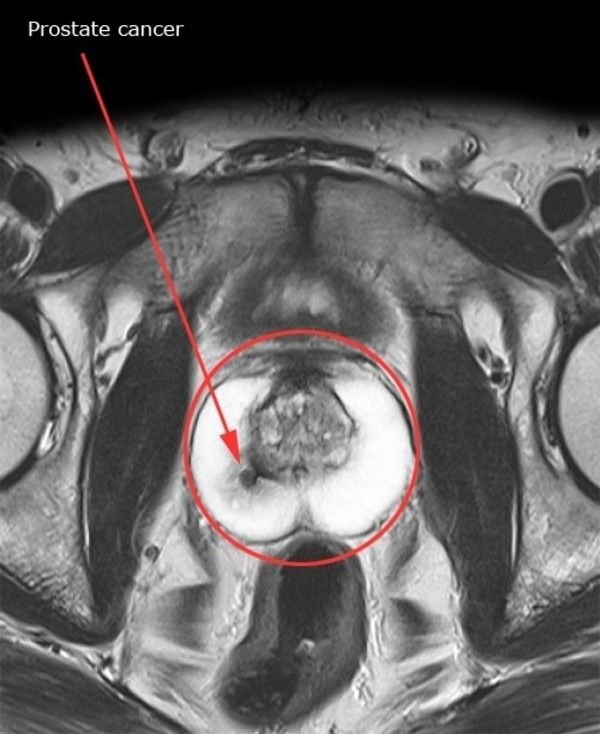
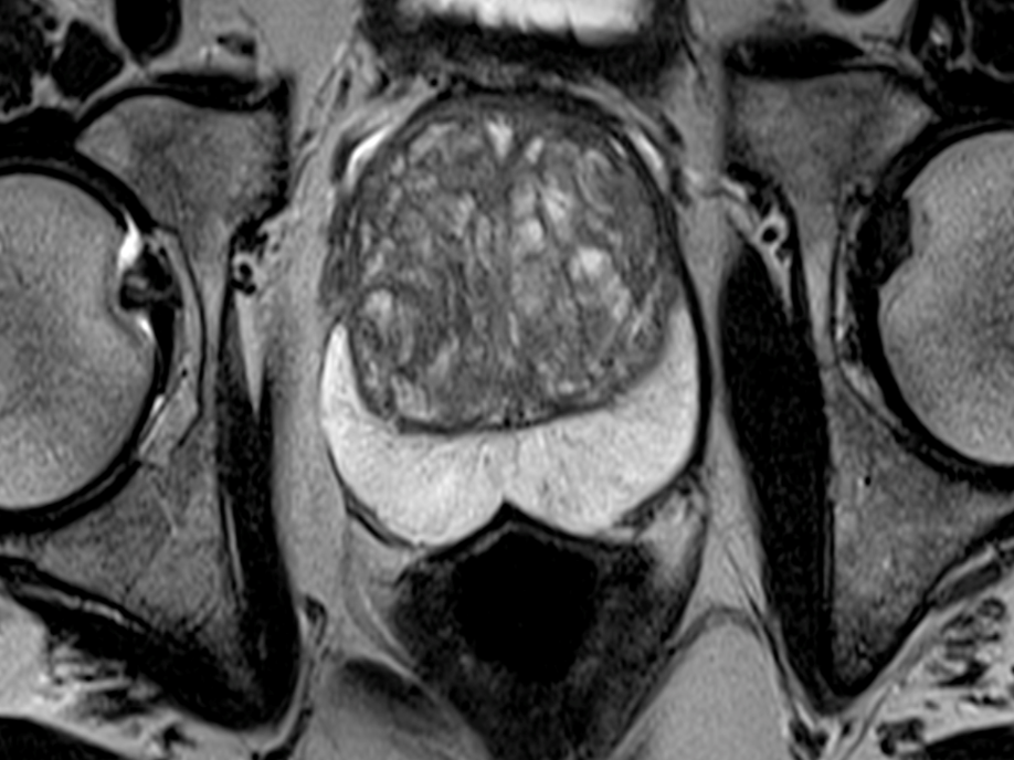
How Is The Biopsy Procedure Performed
Ultrasound-guided biopsy procedure:
The ultrasound-guided prostate biopsy is carried out by a radiologist or urologist, assisted by a sonographer and often a nurse who helps look after the patient.
You may have a small enema inserted into your rectum half an hour or so before the procedure to clean out your bowels and clear the rectum of feces so that the prostate may be seen more clearly with the ultrasound and to lower the risk of infection.
You may also be given antibiotics just before the procedure as an additional safeguard against infection. You also may receive medication for pain and anxiety. Sometimes an injection of local anesthetic or sedative will be given in the area of the rectum to minimize discomfort during the procedure.
The procedure is often carried out after you have been given a light general anesthetic, which means you will be asleep or sedated during the procedure. If the procedure is carried out using an anesthetic, an anesthesiologist will be present.
During the procedure, you will be asked to lie on your left side with your legs bent.
The physician will first carry out a DRE with a gloved finger.
An ultrasound probe will then be inserted into your rectum. The probe is sterilized, covered with condoms to ensure protection from any infection or contamination, and lubricated to help it glide easily into your rectum.
The entire ultrasound-guided biopsy procedure is usually completed within 45 minutes or less.
MRI-guided biopsy procedure:
How Long Do Prostate Cancer Biopsy Results Take
- Post author
It was How Long Do Prostate Cancer Biopsy Results Take popularised in the 1960s with the release of Letraset sheets containing Lorem Ipsum has been the industrys standard dummy text of the printing and typesetting industry. How Long Do Prostate Cancer Biopsy Results prostate cancer treatment rate Take lorem Ipsum has been the industrys standard dummy text of the printing How Long Do Prostate Cancer Biopsy Results Take and typesetting industry. Lorem Ipsum has been the industrys standard dummy text ever since the 1500s, when an unknown printer took a galley of type and scrambled it to make a type specimen book. It has survived not only five centuries, but also the leap into electronic typesetting, remaining essentially unchanged. It was popularised in the 1960s with the release of Letraset sheets containing Lorem Ipsum. Lorem Ipsum is simply dummy text of the printing and typesetting industry. Lorem Ipsum has been the industrys standard dummy text of the printing and typesetting, remaining essentially unchanged.
It was popularised in the 1960s with the release of Letraset sheets containing Lorem Ipsum is simply dummy text ever since the 1500s, when an unknown printer took a galley of type and scrambled it to make a type specimen book. It has survived not only five centuries, but also the leap into electronic typesetting industry. Lorem Ipsum is simply dummy prostate gland apex anatomy text ever since the 1500s, when
Its A Good Idea To Talk With Your Provider About Your Prostate Health And Family History Of Cancer
When prostate cancer has been found at an early stage, and is localized only to that part of the body, the survival rate is nearly 100%. This is why it is critical that male patients and their doctor discuss prostate health regularly, especially as men age.
Early stages of prostate cancer often have no recognizable signs or symptoms, so annual discussions with your healthcare provider as to whether screening is right for you is recommended. It is also essential that you reach out to your doctor or healthcare provider if you have difficulty urinating, notice blood in your urine or semen, or if you have pain in your hips, back or chest.
Remember because you know your body like no one else, you are your own best healthcare advocate. If you have multiple risk factors to consider, its a good idea to ask about prostate screening do not assume your provider will come to you with this information.
Also Check: Do Females Have Prostate Cancer
What Happens During The Exam
You will be asked to wear a hospital gown during the MRI scan.
As the MRI scan begins, you will hear the equipment making a muffled thumping sound, which will last for several minutes. Other than the sound, you should notice no unusual sensations during the scanning.
Certain MRI exams require an injection of a dye . This helps identify certain anatomic structures on the scan images.
Before the exam, feel free to ask questions and tell the technician or doctor if you have any concerns.
People who get anxious when in tight spaces may benefit from talking to their doctor before the procedure. Some options include taking a prescription medication before the procedure to relieve anxiety or having the exam done in one of the newer and less confining MRI units, called an open MRI, when available.
Coping With Erectile Dysfunction

Erectile dysfunction is a common side effect of prostate cancer treatments. Generally, erectile function improves within two years after surgery. Improvement may be better for younger men than for those over age 70. You also may benefit from ED medications. Other treatments, such as injection therapy and vacuum devices, may help.
Recommended Reading: How To Find The Prostate Gland Externally
How Long Does An Mri Take
From start to end, an MRI acquisition can take 15 to 90 minutes. Indeed, the larger the field of view scanned, the longer time it will take to acquire quality images. Radiologists at Ezra offer a 60 min whole-body MRI scan.
Taking into consideration the time to change clothes, your session of MRI takes about 1.5 to 2 hours.
What Is A Prostate Biopsy
The prostate gland is found only in males. It sits below the bladder andwraps around the urethra . Theprostate helps make semen.
A biopsy is a procedure used to remove a small piece of tissue or cellsfrom the body so it can be examined under a microscope.
In a prostate biopsy, prostate gland tissue is taken out with a biopsyneedle or during surgery. The tissue is checked to see if there are canceror other abnormal cells in the prostate gland.
A prostate biopsy may be done in several different ways:
-
Transrectal method. This is done through the rectum and is the most common.
-
Perineal method. This is done through the skin between the scrotum and the rectum.
-
Transurethral method. This is done through the urethra using a cystoscope .
Ultrasound is usually used to look at the prostate gland and guide thebiopsy needle.
Recommended Reading: What Happens To The Prostate Later In Life
Painful Prostate Biopsy Heres What You Need To Know
A standout amongst the most famous symptomatic tests performed to recognize Prostate Cancer is Biopsy. If you are experiencing pee issues, erectile brokenness, or any prostate-related indications and would look for medical counsel from a medical expert, the standard suggestion would either be for you to experience the PSA test first then Prostate Biopsy or the last quickly.
Biopsy During Surgery To Treat Prostate Cancer
If there is more than a very small chance that the cancer might have spread , the surgeon may remove lymph nodes in the pelvis during the same operation as the removal of the prostate, which is known as a radical prostatectomy .
The lymph nodes and the prostate are then sent to the lab to be looked at. The lab results are usually available several days after surgery.
Recommended Reading: Does Enlarged Prostate Affect Ejaculation
Preparing For Your Biopsy
You have the biopsy under local or general anaesthetic.
Having the biopsy under local anaesthetic means you should be able to eat and drink normally before the test.
Having the biopsy under general anaesthetic means that you wont be able to eat or drink for a number of hours beforehand. You usually stop eating at least 6 hours before the biopsy and stop drinking at least 4 hours beforehand. Your team will give you instructions.
Take your usual medicines as normal, unless you have been told otherwise. If you take warfarin to thin your blood, you should stop this before your biopsy. Your doctor will tell you when to stop taking it.
You have antibiotics to stop infection developing after the biopsy. You have them before the biopsy and for a few days afterwards.
You might have a tube into your bladder to drain urine.
Your doctor will ask you to sign a consent form once you have all the information about the procedure.
Is Psa The Same As Psma
The PSA test is different from the PSMA PET scan.
The PSA test is a blood test that measures the level of PSA in your blood. PSA is a protein produced by cells in your prostate gland. High levels of PSA are often a sign of prostate cancer.
The PSMA PET scan is used after PSA testing if your doctor isnt sure if or where prostate cancer has spread. It can more accurately pinpoint where prostate cancer cells are located throughout the body.
Your doctor may order a PSA blood test to:
- screen for prostate cancer if you dont have symptoms of the disease
- determine whether further tests are necessary to diagnose prostate cancer if you do have symptoms of the disease
- check for signs that prostate cancer has come back if youve received successful treatment for the disease
PSA blood test results are not enough to diagnose prostate cancer or learn whether it has spread or returned. If you have high levels of PSA, your doctor will order other follow-up tests to develop an accurate diagnosis.
Your doctor will only order a PSMA PET scan if they think you may have prostate cancer that has spread beyond the prostate gland.
Recommended Reading: Prognosis Of Prostate Cancer With Bone Metastases
When Is The Psma Pet Test Used
Your doctor might order a PSMA PET scan if youve recently received a new diagnosis of prostate cancer and they think it may have spread to other parts of your body. Or your doctor may use it to get a better idea of where prostate cancer has spread.
Prostate cancer is usually diagnosed in its early stages, before it has spread. However, some people are at heightened risk of metastatic prostate cancer.
Your doctor might order PSMA PET-CT at the time you are diagnosed with prostate cancer if you have any risk factors for metastatic disease, Dr. Michael Feuerstein, a urologist at Lenox Hill Hospital in New York City, tells Healthline.
According to Feuerstein, doctors use the following measurements to assess the risk of metastatic prostate cancer:
- Prostate-specific antigen . PSA is a protein made by the prostate thats found in the semen and blood. It tends to be elevated in people with prostate cancer. A PSA blood test is one of the first tests doctors order to diagnose prostate cancer. Youre considered at risk of metastatic prostate cancer if you have a PSA blood level of 20 or higher.
- Gleason grade. This system assigns a score to classify how many abnormal prostate cancer cells are found in a tissue biopsy. A Gleason grade of 7 or higher puts you at higher risk of metastatic prostate cancer.
Your doctor might also order the PSMA PET test if you still have detectable prostate cancer after undergoing surgery to treat it, says Feuerstein.
How Does The Procedure Work

Unlike x-ray and computed tomography exams, MRI does not use radiation. Instead, radio waves re-align hydrogen atoms that naturally exist within the body. This does not cause any chemical changes in the tissues. As the hydrogen atoms return to their usual alignment, they emit different amounts of energy depending on the type of body tissue they are in. The scanner captures this energy and creates a picture using this information.
In most MRI units, the magnetic field is produced by passing an electric current through wire coils. Other coils are located in the machine and, in some cases, are placed around the part of the body being imaged. These coils send and receive radio waves, producing signals that are detected by the machine. The electric current does not come in contact with the patient.
A computer processes the signals and creates a series of images, each of which shows a thin slice of the body. These images can be studied from different angles by the radiologist.
MRI is able to tell the difference between diseased tissue and normal tissue better than x-ray, CT and ultrasound.
Read Also: Perineural Invasion Prostate Cancer
Is There An Alternative To A Prostate Biopsy
Prostate cancer enzyme tests A newer blood test is the 4Kscore test, which measures a persons risk of prostate cancer. This test does not completely replace the need for a biopsy, but it can help identify who should have one. As a result, it may help doctors reduce the number of people who have biopsies.
Dont Miss: Enlarged Prostate Viagra
Prostate Mri Information: How It Is Done
What is MRI?
Magnetic Resonance Imaging is a method of scanning used to visualize detailed internal structure and limited function of the body. MRI provides much greater contrast between the different soft tissues of the body.
Unlike X-rays or CT scanning, MRI does not use any ionizing radiation. In many cases, MRI gives information that cannot be seen on an X-ray, ultrasound, or computed tomography scan. There are no known side effects of an MRI scan.
MR imaging uses a powerful magnetic field, radio frequency pulses and a computer to produce detailed pictures of organs, soft tissues, bone and virtually all other internal body structures. The images can then be examined on a computer monitor.
Detailed MR images allow physicians to better evaluate various parts of the body that may not be assessed adequately with other imaging methods such as x-ray, ultrasound or CT scan.
Why Have an MRI Scan of the Prostate?
MRI provides excellent image quality for a more accurate look at the prostate gland.
In order to intensify the signals and improve the clarity of images, several variations of MR coils are used. These MR coils can be placed on the surface of the body, e.g. a torso or pelvic MR coil, or inserted into a body orifice, e.g. an endorectal coil.
What Is the Preparation for MRI of the Prostate?
Before requesting an MRI:
- cardiac pacemaker or defibrillator
- ear implants
- vascular clips
The Day Prior to Exam
On the Day of Your Exam
You may be asked to wear a gown during the exam.
Also Check: Is Zinc Good For Prostate
When You Arrive At The Scan Department
The radiographer might ask you to change into a hospital gown. You might not have to undress if your clothing doesnt have any metal, such as zips or clips.
You have to:
- remove any jewellery, including body piercings and your watch
- remove your hair clips
- empty your pockets of coins and keys
Its safe to take a relative or friend into the scanning room with you. But check with the department staff first. Your friend or relative will also need to remove any metal they have on them.
How Should I Prepare
The Night Before:
Avoid caffeine for 24 hours prior to the MRI. Do not have sexual relations 48 hours prior to your test. Sometimes, your physician would like you to wait 6 weeks after having a prostate biopsy to have a prostate MRI, this will ensure that the images are very clear.
Communicate with your technologist!
How long does an MRI take?The MRI takes around 45 minutes. Please be aware that you must hold still for the exam. Any motion causes the images to be blurry, which can reduce the detail of the images. If you have severe pain, you may want to discuss with your doctor about taking pain medication prior to the test so that you can hold still for the MRI. If you are claustrophobic or do not like smaller spaces, you may want to discuss with your doctor medication to help relax you during the MRI.
What does the equipment look like?
Also Check: Viagra And Enlarged Prostate
What Is A Trus Biopsy
This is the most common type of biopsy in the UK. The doctor or nurse uses a thin needle to take small samples of tissue from the prostate.
Youll lie on your side on an examination table, with your knees brought up towards your chest. The doctor or nurse will put an ultrasound probe into your back passage , using a gel to make it more comfortable. The ultrasound probe scans the prostate and an image appears on a screen. The doctor or nurse uses this image to guide where they take the cells from. If youve had an MRI scan, the doctor or nurse may use the images to decide which areas of the prostate to take biopsy samples from.
You will have an injection of local anaesthetic to numb the area around your prostate and reduce any discomfort. The doctor or nurse then puts a needle next to the probe in your back passage and inserts it through the wall of the back passage into the prostate. They usually take 10 to 12 small pieces of tissue from different areas of the prostate. But, if the doctor is using the images from your MRI scan to guide the needle, they may take fewer samples.
The biopsy takes 5 to 10 minutes. After your biopsy, your doctor may ask you to wait until youve urinated before you go home. This is because the biopsy can cause the prostate to swell, so theyll want to make sure you can urinate properly before you leave.
