Tests To Diagnose And Stage Prostate Cancer
Most prostate cancers are first found as a result of screening. Early prostate cancers usually dont cause symptoms, but more advanced cancers are sometimes first found because of symptoms they cause.
If prostate cancer is suspected based on results of screening tests or symptoms, tests will be needed to be sure. If youre seeing your primary care doctor, you might be referred to a urologist, a doctor who treats cancers of the genital and urinary tract, including the prostate.
The actual diagnosis of prostate cancer can only be made with a prostate biopsy .
On this page
Biopsy During Surgery To Treat Prostate Cancer
If there is more than a very small chance that the cancer might have spread , the surgeon may remove lymph nodes in the pelvis during the same operation as the removal of the prostate, which is known as a radical prostatectomy .
The lymph nodes and the prostate are then sent to the lab to be looked at. The lab results are usually available several days after surgery.
You May Be Asked To Drink Some Contrast Material That Moves Through Your Stomach And Bowel
A fludeoxyglucose f 18 injection.* pet/ct scan of the brain was performed on biograph vision. The total time it takes for a psma pet scan is about two hours. It seems that this test is the most advanced modern method to detect undetectable prostate membrane cells in my body. The study shows normal uptake in brain with excellent delineation of functioning cortex and sharp contrast between cortex and white matter. If you are having an fdg pet scan, you will be asked to rest quietly in a bed or arm chair, avoiding movement or talking for 90 minutes. During this time you will be alone as there is limited room for visitors, and it will prevent your friends or relatives from receiving unnecessary radiation exposure. You may be asked to drink some contrast material that moves through your stomach and bowel. Additionally there is sharp basal ganglial edge definition, especially the sharp margins and distinct separation of the head of caudate nucleus and. What will happen is the tracer will be. Improved diagnosis and new medications may lead to read more. I’ve looked at 6+ discussions, webinars, videos, lectures about this effectiveness of this pet/ct scan. 13.06.2019 · i’m getting a psma pet/ct total body scan scheduled as soon as possible. What to expect from proton therapy.
Also Check: What Is The Definition Of Prostate Gland
Greater Accuracy And Changing Treatment
Approximately 300 men were enrolled in the Australian trial, all with newly diagnosed localized prostate cancer , and all were considered to have high-risk disease. For all men in the trial, the planned treatment was either surgery or radiation therapy to the prostate only.
Half the men were randomly assigned to initially undergo a CT and bone scan, and the other half to PSMA PET-CT.
Based on the imaging, PSMA PET-CT was 27% more accurate than the standard approach at detecting any metastases . Accuracy was determined by combining the scans sensitivity and specificity, measures that show a tests ability to correctly identify when disease is present and not present.
PSMA PET-CT was more accurate for both metastases found in lymph nodes in the pelvis and in more distant parts of the body, including bone. Radiation exposure was also substantially lower with PSMA PET-CT than with the conventional approach.
The trial investigators also tracked how imaging results influenced clinicians treatment choices. Based on imaging findings, the initial treatment plan was changed for 15% of men who underwent conventional imaging compared with 28% of men who underwent PSMA PET-CT.
Another key finding, Dr. Hofman noted, was that PSMA PET-CT was much less likely to produce inconclusive, or equivocal, results .
Thats important, he continued, because if you have a scan with equivocal findings, it often leads to more scans or biopsies or other tests.
General Example Of Pet Scan Use After A Prostate Cancer Diagnosis

It is important to note that while PET Scanning can be an essential tool in the assessment of prostate cancer, it is not always used on all patients, and there are many other imaging tests and procedures that may be recommended depending on the patients specific needs.
This is an example of a prostate cancer care that would include PET scanning.
You May Like: Cranberry Juice Prostate
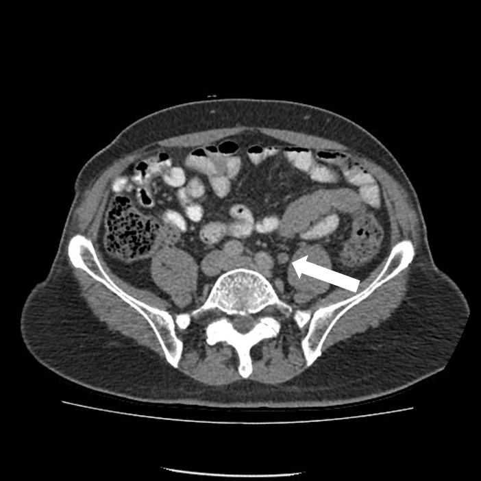
You Might Have A Ct Scan Of Your Head And Neck Your Chest Or Your Abdomen
It may grow slowly and it’s typically treatable. Find out what it is and how you have it. Prostate cancer is a common type of cancer in men, according to the mayo clinic. One in seven men in the united states will receive a prostate cancer diagnosis during his lifetime. A ct scan can check if testicular cancer has spread to any lymph nodes in the tummy .
This diagnostic tool is used in many different medical situations, as it gives doctors a way to visualize the body internally to determine the e. You may have a ct scan to stage for vulval cancer. Although screenings for prostate cancer are one tool for early detecti. If your doctor orders a computerized tomography scan, you’ll be preparing to undergo a ct scan. Being armed with information is vital to begin the fight.
Lymph Node Biopsy As A Separate Procedure
A lymph node biopsy is rarely done as a separate procedure. Its sometimes used when a radical prostatectomy isnt planned , but when its still important to know if the lymph nodes contain cancer.
Most often, this is done as a needle biopsy. To do this, the doctor uses an image to guide a long, hollow needle through the skin in the lower abdomen and into an enlarged node. The skin is numbed with local anesthesia before the needle is inserted to take a small tissue sample. The sample is then sent to the lab and looked at for cancer cells.
Read Also: Zinc And Prostate
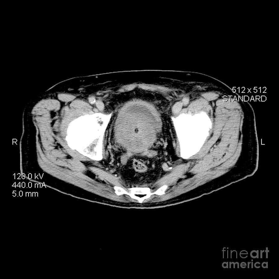
A Negative Ct Scan For Prostate Cancer Isnt Anything To Get Too Hopeful About Because A Ct Scan Can Actually Miss Prostate Cancer
Yes, a CT scan can miss prostate cancer, says Jonathan W. Simons, MD, President and Chief Executive Officer of the Prostate Cancer Foundation, David H. Koch Chair.
Dr. Simons explains, A CT scan is fundamentally a three-dimensional X-ray of the body. It does not intrinsically distinguish between cancer and normal tissue.
We therefore rely on anatomical changes to tell us the probability something is normal or is cancer.
For example, if a lymph node is normal in size it still has the potential to have cancer in it.
However, if a lymph node is enlarged it has a much higher probability there is cancer present, although not guaranteed.
Any imaging modality cant identify microscopic disease, so a CT scan, MRI scan and PET scan can miss prostate cancer that may have spread.
The presence of prostate cancer can be indicated by an abnormally elevated PSA blood test result or by a digital rectal exam .
These tests do not diagnose a malignancy they can only raise suspicion. Diagnosis is made only via a biopsy of tissue extracted from the prostate gland.
The Study Shows Normal Uptake In Brain With Excellent Delineation Of Functioning Cortex And Sharp Contrast Between Cortex And White Matter
What to expect from proton therapy. If you are having an fdg pet scan, you will be asked to rest quietly in a bed or arm chair, avoiding movement or talking for 90 minutes. 13.06.2019 · i’m getting a psma pet/ct total body scan scheduled as soon as possible. It seems that this test is the most advanced modern method to detect undetectable prostate membrane cells in my body. I’ve looked at 6+ discussions, webinars, videos, lectures about this effectiveness of this pet/ct scan. You may be asked to drink some contrast material that moves through your stomach and bowel. A fludeoxyglucose f 18 injection.* pet/ct scan of the brain was performed on biograph vision. Once the psma pet scan is completed, men can receive prostate cancer care at ucla health’s hospitals in westwood and santa monica, or at ucla health’s encino medical office. The scan can also be used in conjunction with care at any other hospital where the patient is being treated. Additionally there is sharp basal ganglial edge definition, especially the sharp margins and distinct separation of the head of caudate nucleus and. Improved diagnosis and new medications may lead to read more. During this time you will be alone as there is limited room for visitors, and it will prevent your friends or relatives from receiving unnecessary radiation exposure. The study shows normal uptake in brain with excellent delineation of functioning cortex and sharp contrast between cortex and white matter.
You May Like: Does Prostatitis Go Away On Its Own
Axumin Pet Scanning For Prostate Cancer Care
Axumin is an FDA-approved agent used for Axumin PET scans for prostate cancer. Axumin is often able to image and restage recurrent prostate cancer better than any other conventional imaging techniques. Biochemical recurrence, typically suspected with rising PSA levels, is the standard in monitoring patients for suspected recurrent prostate cancer. Traditional imaging techniques are often limited in that they may detect a small lymph node or suspicious finding, but cannot further functionally characterize the molecular activity to determine the level of suspicion. The introduction of Axumin PET scanning has been a breakthrough, allowing physicians the ability to accurately locate and restage prostate cancer with precision, especially in the setting of suspiciously rising PSA levels.
How Do Axumin PET Scans Work?
An Axumin PET uses a radioactive tracer, given as an injection, that is linked to an amino acid which is absorbed by prostate cancer at a much more rapid rate than normal cells. The rapid uptake of Axumin by prostate cancer cells is then imaged by the advanced technology within the PET scan equipment. The PET scan images are then reviewed in order to determine if there has been any spread to other areas in the body.
Preparing For Your Pet
For most PET-CT scans, you need to stop eating about 4 to 6 hours beforehand. You can usually drink water during this time. You might have instructions not to do any strenuous exercise for 24 hours before the scan.
Some people feel claustrophobic when theyre having a scan. Contact the department staff before your test if youre likely to feel like this. They can take extra care to make sure youre comfortable and that you understand whats going on.
Your doctor can arrange to give you medicine to help you relax, if needed.
Recommended Reading: What Is The Va Disability Rating For Prostate Cancer
Psma Pet Scanning For Prostate Cancer Care
PSMA PET scans are now FDA-approved and ongoing studies continue to suggest that it will be an even more valuable imaging tool used for both staging and restaging prostate cancer in the near future. The specifics of PSMA will hopefully allow for even more accurate imaging and the earliest possible detection when performing PET scans for prostate cancer.
When Is A PSMA PET Scan Used For Prostate Cancer?
Now FDA approved, we are currently performing PSMA PET Scans at our facility for the following purposes.
- Staging: Patients with a first-time diagnosis of prostate cancer benefit from using a PSMA PET scan because of its superior ability to detect cancer spread offering the best possible future treatment options.
- Restaging: Patients with suspected biochemically recurrent prostate cancer, meaning patients with elevated PSA levels after definitive therapy, benefit from using PSMA PET scans because of its ability to precisely locate recurrent prostate cancer and detect cancer spread. For both of these reasons, PSMA PET scans offer the best treatment options for recurrent prostate cancer.
How Do PSMA PET Scans Work?
Recommended Reading: Can Prostate Cancer Go Away
Prostate Cancer Urine Test

This test detects the gene PCA3 in your urine and can also help your doctorbetter assess your prostate cancer risk.
PCA3 is a prostate-specific noncoding RNA. Its a gene thats only in yourprostate. If the gene is overexpressed , then theres a greater chance you have prostate cancer.
Like PSA and PHI tests, this isnt definitive, either. But data suggestthat when cancer is present, the PCA3 will be positive 80 percent of thetime. This test can also help your doctor determine whether a biopsy isnecessary.
Both of these new tests are more accurate than the PSA test. Your doctormay recommend one or more than one, based on the specifics of your case.
Read Also: Viagra Bph
Recommended Reading: Does Enlarged Prostate Affect Ejaculation
Determining Whether Prostate Cancer Is Aggressive
If a biopsy sample is found to contain cancer, the pathologist analyzing the specimen takes a deeper look at the cancer cells to determine how aggressive the disease is likely to be.
If the cancer cells appear significantly abnormal and dissimilar from healthy cells under a microscope, the cancer is considered more aggressive and expected to advance quickly. Conversely, cancer cells that look relatively similar to healthy cells indicate that its less aggressive and may not spread as fast.
Prostate cancers are assigned a Gleason score depending on how abnormal the cells look..
Gleason score: Gleason scores range from 2 to 10, going from least to most aggressive prostate cancers.
There are different types of cancer cells in a prostate tumor, so the final Gleason score is determined by adding the scores of the two main areas of the tumor.
First, the primary part of the tumor is assigned a number between 1 and 5. Lower numbers indicate that the cells appear relatively similar to healthy cells, while higher numbers show that the cells are abnormal-looking. Then, another number between 1 and 5 is assigned to describe the second most prevalent area of the tumor.
Finally, the two numbers assigned to the different parts of the prostate tumor are added. So, if most of the tumor is given a 4, and some of the tumor is more aggressive and given a 5, the final Gleason score would be 9.
There are many biomarker tests, including:
- Oncotype DX® Genomic Prostate Score
- Prolaris
- ProMark®
A Note On Suspicious Results
A suspicious result indicates that the biopsy sample contained some abnormalities but no cancer was found. There are a couple of potential explanations for a suspicious prostate biopsy result, including:
- Prostatic intraepithelial neoplasia refers to changes within prostate cells that are abnormal, but not indicative of cancer. This condition is low-grade or high-grade, depending on how abnormal the cells are. Low-grade PIN is very common and isn’t associated with prostate cancer. High-grade PIN, however, is associated with a higher risk of prostate cancer. If you have high-grade PIN after a prostate biopsy, your doctor may recommend that biomarker tests be performed on the sample to learn more about the cells. Alternatively, another prostate biopsy may be suggested.
- Atypical small acinar proliferation indicates that the biopsy sample contains some cells that appear to be cancerous, but not enough to confirm the diagnosis. In most cases, this finding suggests that another prostate biopsy is needed.
- Proliferative inflammatory atrophy describes a prostate biopsy that reveals inflammation in the prostate and abnormally small prostate cells. While these cells arent cancerous, having PIA may be associated with an increased risk of developing prostate cancer.
Read Also: Is Zinc Good For Prostate
How Is Prostate Cancer Diagnosed And Evaluated
Your primary doctor will ask about your medical history, risk factors and symptoms. You will also undergo a physical exam.
Many patients undergo regular prostate cancer screening before symptoms appear. Screening may involve one or more of the following tests:
- Prostate-specific antigen : This test analyzes a blood sample for levels of PSA, a protein the prostate produces. Higher PSA levels could indicate cancer is present.
- Digital Rectal Exam :This test examines the lower rectum and the prostate gland to check for abnormalities in size, shape or texture. The term “digital” refers to the doctor’s use of a gloved, lubricated finger to conduct the exam.
If screening test results are abnormal, your doctor may perform the following imaging tests:
How Do Doctors Decide Which Imaging A Person Should Receive
We usually use CT first for most people, unless a tumor is much better seen on MRI. But we go back and forth as needed. If we see something on a CT scan were unsure about, we may recommend an MRI for further evaluation. If someone has several MRIs and is unable to lie still or hold their breath so we can get a good image, we may suggest a CT as an alternative. Were guided by the principle of whether the benefits of a test outweigh its risks. Thats what medical imaging is about.
Don’t Miss: What Is The Definition Of Prostate Gland
